. 2015 Apr 2:6:6411.
doi: 10.1038/ncomms7411.

Sox2 antagonizes the Hippo pathway to maintain stemness in cancer cells

Affiliations

Sox2 antagonizes the Hippo pathway to maintain stemness in cancer cells

Upal Basu-Roy et al. Nat Commun. .

Abstract

The repressive Hippo pathway has a profound tumour suppressive role in cancer by restraining the growth-promoting function of the transcriptional coactivator, YAP. We previously showed that the stem cell transcription factor Sox2 maintains cancer stem cells (CSCs) in osteosarcomas. We now report that in these tumours, Sox2 antagonizes the Hippo pathway by direct repression of two Hippo activators, Nf2 (Merlin) and WWC1 (Kibra), leading to exaggerated YAP function. Repression of Nf2, WWC1 and high YAP expression marks the CSC fraction of the tumor population, while the more differentiated fraction has high Nf2, high WWC1 and reduced YAP expression. YAP depletion sharply reduces CSCs and tumorigenicity of osteosarcomas. Thus, Sox2 interferes with the tumour-suppressive Hippo pathway to maintain CSCs in osteosarcomas. This Sox2-Hippo axis is conserved in other Sox2-dependent cancers such as glioblastomas. Disruption of YAP transcriptional activity could be a therapeutic strategy for Sox2-dependent tumours.


Figures

Figure 1
Figure 1. Gene expression analysis of Sox2-regulated genes in osteosarcomas
(a) Heat map of hierarchically clustered genes in mOS-482 cells expressing scrambled (Scr) or Sox2 shRNA-A (shSox2-A). The expression level of all 22,670 entities is shown in triplicate samples. Significantly upregulated (red) and downregulated (blue) GO terms and enrichment P values by DAVID are shown. (b) Venn diagram showing intersection of differentially upregulated genes in Sox2-depleted mOS-379 and mOS-482 cells. Nf2 and WWC1 were identified in the overlap. (c) qRT-PCR of mRNA expression of YAP-TEAD transcriptional target genes in Sox2-depleted mOS-482 cells using two independent Sox2 shRNAs (shSox2-A and shSox2-B). Gene expression was normalized relative to actin. *P<0.05 by ANOVA, Error bars, mean ± s.d. western blot of Sox2 expression is shown.
Figure 2
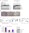
Figure 2. Deregulation of the Hippo pathway in osteosarcomas
(a) Western analysis of YAP, Nf2 and WWC1 in murine and human osteosarcoma cell lines. (b) YAP and Sox2 expression in a spontaneous osteosarcoma in a bone-specific p53/Rb-knockout mouse. Massie's trichrome stain (left panel) and immunohistochemistry with Sox2 (middle panel) and YAP (right panel) antibodies demonstrate that differentiated areas of the tumour (stained blue for collagen) have low expression of Sox2 and YAP. Scale bar, 100 μm. (c) mRNA expression of YAP, Nf2 and WWC1 in human osteosarcomas tumour samples. Box plot shows mRNA expression of YAP, Nf2 and WWC1, as compared with control (red line). A=chondroblastic osteosarcoma; B =fibroblastic osteosarcoma; C=osteoblastic osteosarcoma; and D=telangietactic osteosarcoma. Data used in this analysis were downloaded from www.oncomine.org. (d) Expression of the 8X-GTIIC(TEAD) luciferase reporter in mOS-482 cells expressing either scrambled or two independent Sox2 shRNAs -A and B. Cells were transfected with a TEAD-Luciferase (Firefly) reporter. Luciferase activity was normalized to Renilla luciferase. *P<0.05 by ANOVA. Error bars represent mean ± s.d. (e) Western analysis of phospho- and total YAP in mOS-482 cells expressing scrambled or two independent Sox2 shRNAs-A and B. The anti-phospho YAP antibody recognizes the phosphorylated S127 residue.
Figure 3
Figure 3. YAP expression marks a population of osteosarcoma stem cells
(a) Western analysis of Sox2, YAP, Nf2 and WWC1 in osteosphere-forming cells and adherent cultures (left panel) and Sca-1High/Sox2High versus Sca-1Low Sox2Low cells (right panel) from mOS-482 cells. (b) Western analysis of YAP in mOS-482 cells expressing scrambled or two independent YAP shRNAs (shRNA-A and shRNA-B). (c) Osteogenic and adipogenic differentiation assay. mOS-482 cells expressing scrambled or YAP shRNA-A/B were plated in either osteogenic or adipogenic medium. Osteogenic differentiation was detected using alkaline phosphatase staining. Adipogenesis was detected by Oil Red O staining of cells induced for 15 days (magnification × 100). Results from representative plates at the indicated times are shown. (d) mOS-482 cells expressing scrambled or YAP shRNAs were grown in increasing doses of methotrexate for 48h and surviving cell populations were counted. IC50 values are shown in the inset.
Figure 4
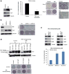
Figure 4. Nf2 is required for osteogenic differentiation of osteosarcomas
(a) Nf2 overexpression in mOS-482 cells increases osteogenic differentiation. Western analysis of Nf2 expression mOS-482 cells transduced with a control or Nf2 retrovirus (left panel). Osteosphere assay (middle panel), and osteogenic and adipogenic differentiation (right panel). *P<0.05 by t-test. Error bars represent mean ± s.d. (b) Nf2 depletion in primary osteoblasts reduces osteogenic differentiation. Analysis of Nf2 in primary osteoblasts transduced with scrambled or two independent shNf2 lentivirus- shNf2-A and shNf2-B. Western analysis (left panel). Osteogenic differentiation (right panel). (c) Depletion of Mst1/2 or Lats1/2 in Nf2-overexpressing cells restores sphere formation. Nf2-overexpressing mOS-482 cells were transfected with control (scrambled) or Mst1/2 siRNAs or Lats1/2 siRNAs. Western analysis (upper panel) and sphere assay (lower panel). *P<0.05 by t-test. Error bars represent mean ± s.d. (d) Control or Nf2-overexpressing mOS-482 cells were transduced with myc-tagged YAP (WT), YAP (5SA—constitutively active YAP mutant) or YAP (94SA—YAP mutant deficient in TEAD-binding) retroviruses. Western analysis (upper panel). Osteogenic differentiation (lower panel).
Figure 5
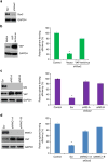
Figure 5. YAP overexpression or Nf2/WWC1 depletion restores osteopheres in Sox2-depleted cells
(a) Western blot showing levels of Sox2 expression in mOS-482–shSox2 cells. (b) Western analysis of YAP expression in shSox2 cells transduced with a control or YAP lentivirus (left panel). Relative sphere-forming efficiency of YAP-transduced shSox2 cells compared with control (parental) mOS-482 cells (right panel). (c) Western analysis of YAP and Nf2 expression in shSox2 cells transfected with scrambled (scr) or two independent Nf2 siRNAs (left panel). Relative sphere-forming efficiency of Nf2-depleted shSox2 cells compared with control (parental) mOS-482 cells (right panel). (d) Western analysis of YAP and WWC1 expression in shSox2 cells transfected with scrambled (scr) or two independent WWc1 siRNAs (left panel). Relative sphere-forming efficiency of WWC1-depleted shSox2 cells compared with control (parental) mOS-482 cells (right panel). *P<0.05 by ANOVA. Error bars represent mean ± s.d.
Figure 6
Figure 6. Sox2 negatively regulates Nf2 by binding to its promoter region
(a) Sox2 binds to the Nf2 5′ region. Sox2 ChIP-Seq shows one peak (green) of Sox2-bound genomic sequence in the Nf2 5′ region in Sox2-overexpressing osteoprogenitors. Inset shows validation of ChIP-Seq in osteosarcoma cells by PCR analysis using primers flanking the Sox2-binding site in the Nf2 5′ region. (b) Analysis of Nf2 expression in mOS-482 cells expressing scrambled or two independent Sox2 shRNAs-A and B. Left panel: Nf2 mRNA expression normalized to actin levels. Middle panel: western analysis of Nf2 expression. Right panel: luciferase assay. A 1.5-kb fragment of the Nf2 promoter containing the Sox2-binding site was cloned into a pGL3-Basic luciferase vector. Activity of the Nf2 5′UTR luciferase reporter construct was normalized to Renilla luciferase activity. *P<0.05 by t-test. Error bars represent mean ± s.d. (c) Analysis of Nf2 expression in primary osteoblasts cells expressing control or Sox2 lentivirus. Left panel—Western analysis of Nf2 expression. Middle panel—Nf2 mRNA expression normalized to actin levels. Right panel—Luciferase assay. Activity of the Nf2 5′UTR luciferase construct was normalized to Renila luciferase activity. *= P<0.05 by t-test. Error bars represent mean ± s.d. (d) Sox2 binding is required for suppression of Nf2 expression. The Nf2 5′ promoter region has one Sox2-binding site. This was mutated in the Nf2 luciferase construct (used in b) and luciferase activity was measured and normalized to Renilla activity. *P<0.05 by t-test. Error bars represent mean±s.d. WT=wild-type promoter, MUT=promoter with a mutant Sox2-binding site. The mutated sites are shown in red in the schematic.
Figure 7
Figure 7. Sox2 negatively regulates WWC1 by binding to its 3′UTR
(a) Sox2 binds to the WWC1 3′UTR. Sox2 ChIP-Seq shows one peak (green) of Sox2-bound genomic sequence in the WWC1 3′UTR in SOX2-overexpressing osteoprogenitors. Inset shows validation of ChIP-Seq in osteosarcoma cells by PCR analysis using primers flanking the Sox2-bidning site in the WWC1 3′UTR. (b) Analysis of WWC1 expression in mOS-482 cells expressing scrambled or two independent Sox2 shRNAs- A and B. Left panel: WWC1 mRNA expression normalized to actin levels. Middle panel: western analysis of WWC1. Right panel: luciferase assay. A 800-bp fragment of the WWC1 3′UTR containing Sox2-binding sites was cloned downstream of the luciferase-coding region in the pTK vector. Activity of the WWC1 3′ UTR luciferase construct was normalized to Renilla luciferase activity. *P<0.05 by t-test. Error bars represent mean ± s.d. (c) Analysis of WWC1 expression in primary osteopblasts cells expressing control or Sox2 lentivirus. Left panel: western analysis of WWC1. Middle panel: WWC1 mRNA expression normalized to actin levels. Right panel: luciferase assay. Activity of the WWC1 3′UTR luciferase construct was normalized to Renila luciferase activity. *P<0.05 by t-test. Error bars represent mean ± s.d.
Figure 8
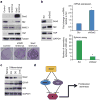
Figure 8. The Sox2-Hippo axis is conserved in multiple cell types
(a) Upper panel: western analysis of YAP, WWC1, Nf2 and Sox2 in NIH-3T3 cells (control, or Nf2 shRNA-A lentivirus or Sox2 lentivirus). Lower panel: colony assay of NIH-3T3 cells expressing the indicated constructs. Colonies were stained with Crystal violet and counted after 10 days. (b) Primary glioblastoma multiforme (GBM) cells were transduced with a control or Sox2 shRNA lentivirus. Left panel: western analysis of Sox2, Nf2 and YAP. Upper right panel—Nf2 mRNA expression normalized to actin. Lower right panel- sphere assay. *P<0.05 by t-test. Error bars represent mean ± s.d. (c) Western analysis of Sox2, GFAP and Nf2 in primary GBM cells grown in sphere cultures (sphere) or differentiated for 3, 6 or 12 days (D3, D6 and D12, respectively). (d) Schematic of Sox2 regulation of Hippo pathway components. Sox2 represses Nf2 and WWC1 that restrain YAP transcriptional function.

Similar articles

Cited by

References

    1. Clarke MF, et al. Cancer stem cells-perspectives on current status and future directions: AACR Workshop on cancer stem cells. Cancer Res. 2006;66:9339–9344. - PubMed
    1. Hanahan D, Weinberg RA. Hallmarks of cancer: the next generation. Cell. 2011;144:646–674. - PubMed
    1. Shackleton M, Quintana E, Fearon ER, Morrison SJ. Heterogeneity in cancer: cancer stem cells versus clonal evolution. Cell. 2009;138:822–829. - PubMed
    1. Clevers H. The cancer stem cell: premises, promises and challenges. Nat Med. 2011;17:313–319. - PubMed
    1. Cordon-Cardo C. Cancer stem cells. Ann Oncol. 2010;21(Suppl 7):vii93–vii94. - PubMed

Publication types

MeSH terms