Review
. 2015 Jul-Aug;6(4):351-67.
doi: 10.1002/wrna.1280. Epub 2015 Apr 1.

PSF: nuclear busy-body or nuclear facilitator?

Affiliations
Review

PSF: nuclear busy-body or nuclear facilitator?

Christopher A Yarosh et al. Wiley Interdiscip Rev RNA. 2015 Jul-Aug.

Abstract

PTB-associated splicing factor (PSF) is an abundant and essential nucleic acid-binding protein that participates in a wide range of gene regulatory processes and cellular response pathways. At the protein level, PSF consists of multiple domains, many of which remain poorly characterized. Although grouped in a family with the proteins p54nrb/NONO and PSPC1 based on sequence homology, PSF contains additional protein sequence not included in other family members. Consistently, PSF has also been implicated in functions not ascribed to p54nrb/NONO or PSPC1. Here, we provide a review of the cellular activities in which PSF has been implicated and what is known regarding the mechanisms by which PSF functions in each case. We propose that the complex domain arrangement of PSF allows for its diversity of function and integration of activities. Finally, we discuss recent evidence that individual activities of PSF can be regulated independently from one another through the activity of domain-specific co-factors.


Figures

Figure 1
Figure 1
Cellular activities of PTB‐associated splicing factor (PSF). A schematic highlighting the cellular activities which PSF has thus far been reported to regulate. Black activities are those for which the mechanism of PSF activity is best understood. Gray activities are those with less experimental support or unclear mechanism. Dotted double arrow indicates coordination of activities through PSF.
Figure 2
Figure 2
Domain structure of PTB‐associated splicing factor (PSF). A schematic of the domains of PSF along the primary sequence of the protein. Numbers indicate amino acid. Domains are as discussed in the text. RGG, RGG box; P, proline‐rich domain including proline/glutamine‐rich subdomain (P,Q); PRL, PR linker; NLS, nuclear‐localization sequence. RRM1, RRM2, NOPS, and coiled‐coil domains are as listed. The portion of PSF that comprises the DBHS core region is noted. Exact amino acid boundaries of the NLS are also given below.
Figure 3
Figure 3
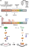
Regulation of PTB‐associated splicing factor (PSF). (a) PSF protein partners. Arrows indicate approximate domain of PSF through which partner proteins interact, in the cases where this is known. Protein partners for which no domain information is available are listed to the top right. (b) Location of mapped sites of phosphorylation on PSF for which the kinase is known. Also shown are additional mapped sites of methylation (Me) and sumoylation (Sumo). (c) Schematic of one pathway of PSF regulation for which details are known. GSK3, PSF, and TRAP‐150 are as described in the text. PKC is protein kinase C, a signaling molecule known to induce inhibitory phosphorylation of GSK3, thereby reducing its activity. Red ‘P’ indicates GSK3‐dependent phosphorylation of PSF. Gray and beige boxes and lines correspond to the exons and introns of the CD45 gene. The central beige exon corresponds to CD45 exon 4, a known target of PSF repressive activity.

References

    1. Patton JG, Porro EB, Galceran J, Tempst P, Nadal‐Ginard B. Cloning and characterization of PSF, a novel pre‐mRNA splicing factor. Genes Dev 1993, 7:393–406. - PubMed
    1. Shav‐Tal Y, Zipori D. PSF and p54(nrb)/NonO—multi‐functional nuclear proteins. FEBS Lett 2002, 531:109–114. - PubMed
    1. Bond CS, Fox AH. Paraspeckles: nuclear bodies built on long noncoding RNA. J Cell Biol 2009, 186:637–644. - PMC - PubMed
    1. Passon DM, Lee M, Rackham O, Stanley WA, Sadowska A, Filipovska A, Fox AH, Bond CS. Structure of the heterodimer of human NONO and paraspeckle protein component 1 and analysis of its role in subnuclear body formation. Proc Natl Acad Sci USA 2012, 109:4846–4850. - PMC - PubMed
    1. Fox AH, Bond CS, Lamond AI. P54nrb forms a heterodimer with PSP1 that localizes to paraspeckles in an RNA‐dependent manner. Mol Biol Cell 2005, 16:5304–5315. - PMC - PubMed

Publication types

Substances