. 2016 May;221(4):2135-46.
doi: 10.1007/s00429-015-1034-7. Epub 2015 Apr 2.

Association of white matter hyperintensities and gray matter volume with cognition in older individuals without cognitive impairment

Affiliations

Association of white matter hyperintensities and gray matter volume with cognition in older individuals without cognitive impairment

Zoe Arvanitakis et al. Brain Struct Funct. 2016 May.

Abstract

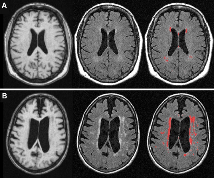
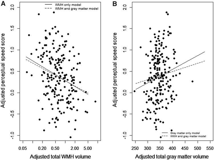
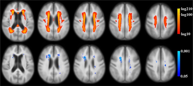
Both presence of white matter hyperintensities (WMH) and smaller total gray matter volume on brain magnetic resonance imaging (MRI) are common findings in old age, and contribute to impaired cognition. We tested whether total WMH volume and gray matter volume had independent associations with cognition in community-dwelling individuals without dementia or mild cognitive impairment (MCI). We used data from participants of the Rush Memory and Aging Project. Brain MRI was available in 209 subjects without dementia or MCI (mean age 80; education = 15 years; 74 % women). WMH and gray matter were automatically segmented, and the total WMH and gray matter volumes were measured. Both MRI-derived measures were normalized by the intracranial volume. Cognitive data included composite measures of five different cognitive domains, based on 19 individual tests. Linear regression analyses, adjusted for age, sex, and education, were used to examine the relationship of logarithmically-transformed total WMH volume and of total gray matter volume to cognition. Larger total WMH volumes were associated with lower levels of perceptual speed (p < 0.001), but not with episodic memory, semantic memory, working memory, or visuospatial abilities (all p > 0.10). Smaller total gray matter volumes were associated with lower levels of perceptual speed (p = 0.013) and episodic memory (p = 0.001), but not with the other three cognitive domains (all p > 0.14). Larger total WMH volume was correlated with smaller total gray matter volume (p < 0.001). In a model with both MRI-derived measures included, the relation of WMH to perceptual speed remained significant (p < 0.001), while gray matter volumes were no longer related (p = 0.14). This study of older community-dwelling individuals without overt cognitive impairment suggests that the association of larger total WMH volume with lower perceptual speed is independent of total gray matter volume. These results help elucidate the pathological processes leading to lower cognitive function in aging.

Keywords: Aging; Brain; Cognition; Gray matter; MRI; Volume; Voxel-wise analyses; White matter hyperintensities.


Figures

Fig. 1
Fig. 1
Brain MRI images from two individuals, one with small (a), and one with large (b) total WMH volume. Top row (a) shows one individual with small (25 %) total WMH volume, and bottom row (b) shows another individual with large (75 %) total WMH volume. The left column shows averaged T 1-weighted MPRAGE images reformatted from sagittal to axial plane, the middle column shows raw axial T 2-weighted FLAIR images, and the right column shows in red the corresponding WMH lesions segmented automatically based on T 1- and T 2-weighted signals
Fig. 2
Fig. 2
Relationships of total WMH and gray matter volumes to perceptual speed. Left panel (a) level of perceptual speed as a function of total WMH volume (on a geometric axis) in a linear regression model without controlling for total gray matter volume (WMH only model) and in a model controlling for total gray matter volume (WMH and gray matter model). Right panel (b) level of perceptual speed as a function of total gray matter volume in a linear regression model without controlling for total WMH volume (gray matter only model) and in a model controlling for total WMH volume (WMH and gray matter model). All models and data values are adjusted for age, sex, and education. WMH volumes were analyzed and adjusted in log-10 scale
Fig. 3
Fig. 3
Relationships of WMH to perceptual speed. Top row maps of the number of participants with WMH in the same location (red–yellow color scale, a logarithmic color scale is used). Voxels with WMH in fewer than 10 participants are not shown in color. Bottom row voxels where presence of WMH was associated with lower perceptual speed score, controlling for age, sex, education, and total gray matter volume (blue color scale)

References

    1. Aggarwal NT, Wilson RS, Bienias JL, De Jager PL, Bennett DA, Evans DA, DeCarli C. The association of magnetic resonance imaging measures with cognitive function in a biracial population sample. Arch Neurol. 2010;67(4):475–482. - PMC - PubMed
    1. Ardekani BA, Guckemus S, Bachman A, Hoptman MJ, Wojtaszek M, Nierenberg J. Quantitative comparison of algorithms for inter-subject registration of 3D volumetric brain MRI scans. J Neurosci Methods. 2005;42:67–76. doi: 10.1016/j.jneumeth.2004.07.014. - DOI - PubMed
    1. Arfanakis K, Fleischman DA, Grisot G, Barth CM, Varentsova A, Morris MC, Barnes LL. Bennett DA (2013) Systemic inflammation in non-demented elderly human subjects: brain microstructure and cognition. PLoS One. 2013;8(8):e73107. doi: 10.1371/journal.pone.0073107. - DOI - PMC - PubMed
    1. Au R, Massaro JM, Wolf PA, Young ME, Beiser A, Seshadri S, D’Agostino RB, DeCarli C. Association of white matter hyperintensity volume with decreased cognitive functioning: the Framingham Heart Study. Arch Neurol. 2006;63(2):246–250. doi: 10.1001/archneur.63.2.246. - DOI - PubMed
    1. Bennett DA, Wilson RS, Schneider JA, Evans DA, Aggarwal NT, Arnold SE, Cochran EJ, Berry-Kravis E, Bienias JL. Apolipoprotein E epsilon4 allele, AD pathology, and the clinical expression of Alzheimer’s disease. Neurology. 2003;60(2):246–252. doi: 10.1212/01.WNL.0000042478.08543.F7. - DOI - PubMed

Publication types

LinkOut - more resources