. 2015 Jun 1;26(11):1995-2004.
doi: 10.1091/mbc.E14-10-1481. Epub 2015 Apr 1.

The Arp2/3 complex has essential roles in vesicle trafficking and transcytosis in the mammalian small intestine

Affiliations

The Arp2/3 complex has essential roles in vesicle trafficking and transcytosis in the mammalian small intestine

Kang Zhou et al. Mol Biol Cell. .

Abstract

The Arp2/3 complex is the only known nucleator of branched F-actin filaments. Work in cultured cells has established a wide array of functions for this complex in controlling cell migration, shape, and adhesion. However, loss of Arp2/3 complex function in tissues has yielded cell type-specific phenotypes. Here we report essential functions of the Arp2/3 complex in the intestinal epithelium. The Arp2/3 complex was dispensable for intestinal development, generation of cortical F-actin, and cell polarity. However, it played essential roles in vesicle trafficking. We found that in the absence of ArpC3, enterocytes had defects in the organization of the endolysosomal system. These defects were physiologically relevant, as transcytosis of IgG was disrupted, lipid absorption was perturbed, and neonatal mice died within days of birth. These data highlight the important roles of the Arp2/3 complex in vesicle trafficking in enterocytes and suggest that defects in cytoplasmic F-actin assembly by the Arp2/3 complex, rather than cortical pools, underlie many of the phenotypes seen in the mutant small intestine.


Figures

FIGURE 1:
FIGURE 1:
Normal intestinal architecture but failure to thrive upon loss of ArpC3 in the intestinal epithelium. (A) Western blots for ArpC3 and actin in extracts prepared from two WT and two ArpC3 cKO pups. (B) WT and ArpC3 cKO littermates at postnatal day 2. (C–F) Relative weights of P0 and P2 pups, as well as the relative intestinal lengths at these two time points. WT weight and intestinal length was set to 1 in each case. n = 7 or 8, p < 0.0001 for both. (G, H) Hematoxylin and eosin staining of neonatal WT and ArpC3 cKO jejuna. Scale bar, 50 μm. (I, J) α-Catenin (green) and nuclei (blue) in WT and ArpC3 cKO jejuna. Scale bar, 20 μm. Dotted lines indicate basement membrane. (K–N) α-Catenin (green) and F-actin (red) in WT and ArpC3 jejuna (K, L) and colon (M, N). Nuclei are blue; dotted lines indicate the basement membrane. Scale bar, 10 μm.
FIGURE 2:
FIGURE 2:
Normal cortical F-actin, polarity, and adhesion in ArpC3 cKO mice. (A–B′) F-actin staining of WT (A) and ArpC3 cKO (B) intestinal sections. A′ and B′ are brightness enhanced to visualize the low levels of lateral F-actin. (C) Quantitation of fluorescence intensity of phalloidin staining of F-actin. (D, E) Transmission electron micrographs of the brush borders of WT and ArpC3 cKO intestine. Scale bar, 2 10 μm. (F, G) Transmission electron micrographs of the apical junctions of WT and ArpC3 cKO intestine. (H, I) Tight junction (ZO-1, green) and adherens junctions (E-cadherin, red) staining in WT and ArpC3 cKO intestine. Scale bar, 10 μm. Dashed line indicates the basement membrane. (J, K) Occludin (red) localization in WT and ArpC3 cKO intestine. (L–O) Whole-mount intestines stained for ZO1. L–N represent the predominant phenotype, and O represents the status of ZO1 in a smaller number of mutant villi. (P, Q) Ezrin (green) and E-cadherin (red) staining of WT and mutant intestine. (R, S) WT and mutant intestinal sections were stained for the trans-Golgi network protein, Grasp64 (red) and F-actin (green). (T, U) Staining for the terminal web and brush border marker myosin 1D (red) in WT and ArpC3 cKO intestine. Scale bars, 10 μm.
FIGURE 3:
FIGURE 3:
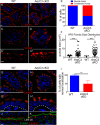
Defects in retromer and lysosomal pools in ArpC3 cKO intestines. WT and ArpC3 cKO intestines were stained with antibodies against the early endosomal marker EEA1 (red) (A,B) or with antibodies against the retromer subunit VPS26 (red) (C–D′). (C′, D′) Magnified views of the apical regions of the intestine. (E) Quantitation of the number of enterocytes with normal/abnormal VPS26 localization. Intestines from WT and mutant animals from two different litters were used for analysis; p = 0.004. (F) Quantitation of the size of VPS26 puncta from two distinct WT and ArpC3 cKO intestines. p = 0.007. (G–H′) Staining for the lysosomal protein Lamp2 (red) in WT and ArpC3 cKO intestinal sections. (G′, H′) Higher-magnification views of the apical localization. Dashed lines indicate basement membranes. (I) Quantitation of cells with normal Lamp2 localization in WT and ArpC3 cKO intestines. Two mice for each genotype; p < 0.01. (J, K) Localization of Lamp2 (red) and F-actin (green) in intestines from neonates taken at late P0. Scale bars, 10 μm, except C′, D′, G′, and H′, 5 μm.
FIGURE 4:
FIGURE 4:
Transcytosis defects in ArpC3 cKO intestines. (A–D) Analysis of endogenous IgGs in WT and ArpC3 cKO intestines, as indicated. (A, B) Neonatal mice that had not suckled; (C, D) mice that had. Sections were stained with donkey anti-mouse antibodies labeled with Rhodamine Red-X to detect the presence of endogenous IgGs. Dashed lines indicate the basement membrane; arrows indicate apical puncta. (E) Quantitation of IgG puncta size. Two mice/genotype, p = 0.0002. (F–I) WT or ArpC3 cKO intestines, as indicated, were incubated with donkey anti–guinea pig-Rhodamine Red-X at 4°C for 15 min and then washed and incubated at 37°C. Samples were fixed after 15 (F, G) and 30 min (H, I), and then imaged. All insets are displayed with apical at the top and basal at the bottom, with the dashed line indicating the basement membrane. Scale bar, 10 μm. Arrows highlight apical puncta of IgG.
FIGURE 5:
FIGURE 5:
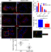
Defect in lipid uptake in the jejunum of the ArpC3 cKO mice. (A, B) Oil Red O staining of WT and ArpC3 cKO mice. Intestinal sections were from the jejunum. (C) Quantitation of sizes of Oil Red O particles in WT and ArpC3 cKO intestine. p = 0.01 for <0.1 μm and 0.03 for 0.1–1.0 μm. D. Quantitation of total intensity of Oil Red O fluorescence in WT and ArpC3 cKO enterocytes. p < 0.01. (E–H) CD36 localization in unfed (E, F) and fed (G, H) intestines. (I) The boxed region in G with F-actin (green) costain. (J) Quantitation of plasma triglyceride levels in WT and ArpC3 cKO neonates (p < 0.05).

Similar articles

Cited by

References

    1. Bernadskaya YY, Patel FB, Hsu HT, Soto MC. Arp2/3 promotes junction formation and maintenance in the Caenorhabditis elegans intestine by regulating membrane association of apical proteins. Mol Biol Cell. 2011;22:2886–2899. - PMC - PubMed
    1. Burd C, Cullen PJ. Retromer: a master conductor of endosome sorting. Cold Spring Harb Perspect Biol. 2014:6, a016774. - PMC - PubMed
    1. Chevrot M, Martin C, Passilly-Degrace P, Besnard P. Role of CD36 in oral and postoral sensing of lipids. Handbk Exp Pharmacol. 2012;2012:295–307. - PubMed
    1. Collins RF, Touret N, Kuwata H, Tandon NN, Grinstein S, Trimble WS. Uptake of oxidized low density lipoprotein by CD36 occurs by an actin-dependent pathway distinct from macropinocytosis. J Biol Chem. 2009;284:30288–30297. - PMC - PubMed
    1. Derivery E, Sousa C, Gautier JJ, Lombard B, Loew D, Gautreau A. The Arp2/3 activator WASH controls the fission of endosomes through a large multiprotein complex. Dev Cell. 2009;17:712–723. - PubMed

Publication types

MeSH terms