. 2015 Jun 1;26(11):2005-19.
doi: 10.1091/mbc.E14-11-1561. Epub 2015 Apr 1.

Centriolar satellite- and hMsd1/SSX2IP-dependent microtubule anchoring is critical for centriole assembly

Affiliations

Centriolar satellite- and hMsd1/SSX2IP-dependent microtubule anchoring is critical for centriole assembly

Akiko Hori et al. Mol Biol Cell. .

Abstract

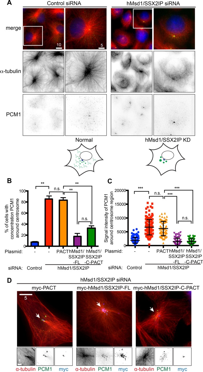
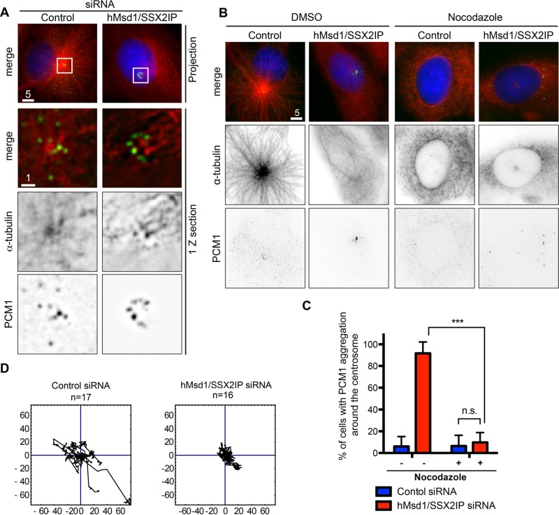
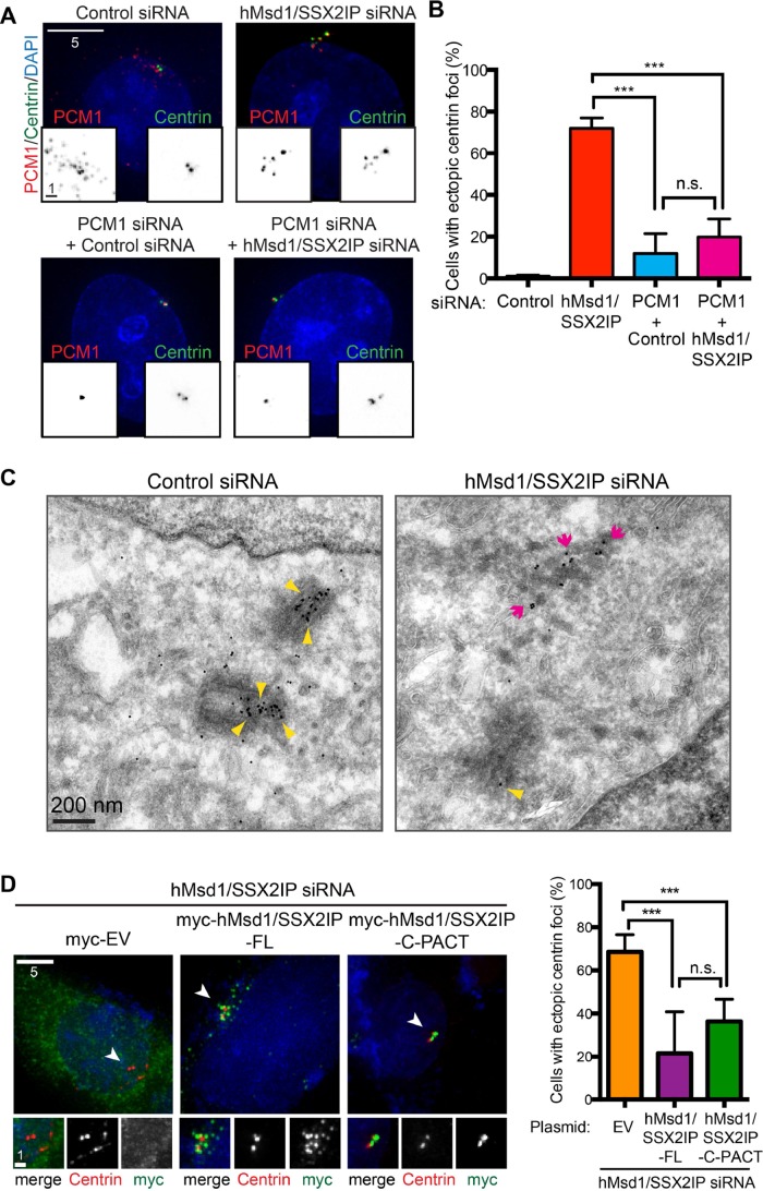
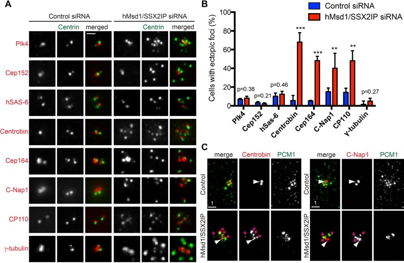
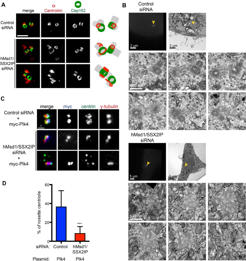
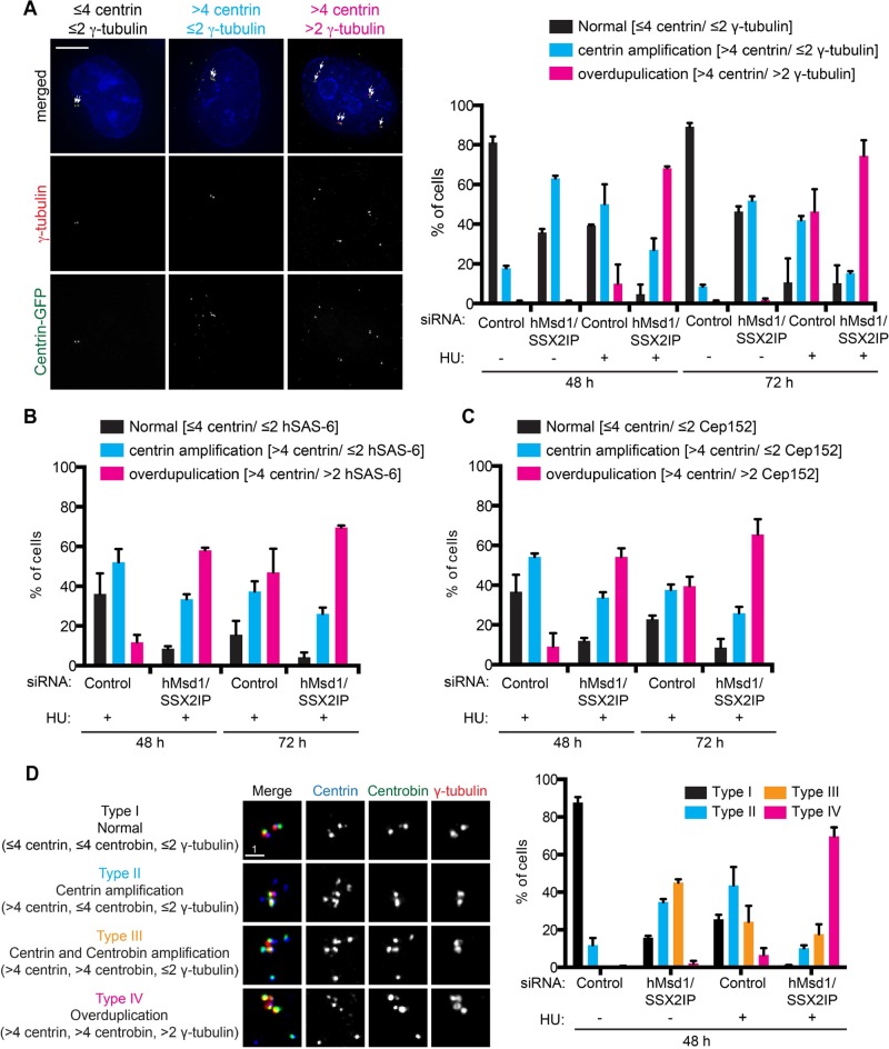
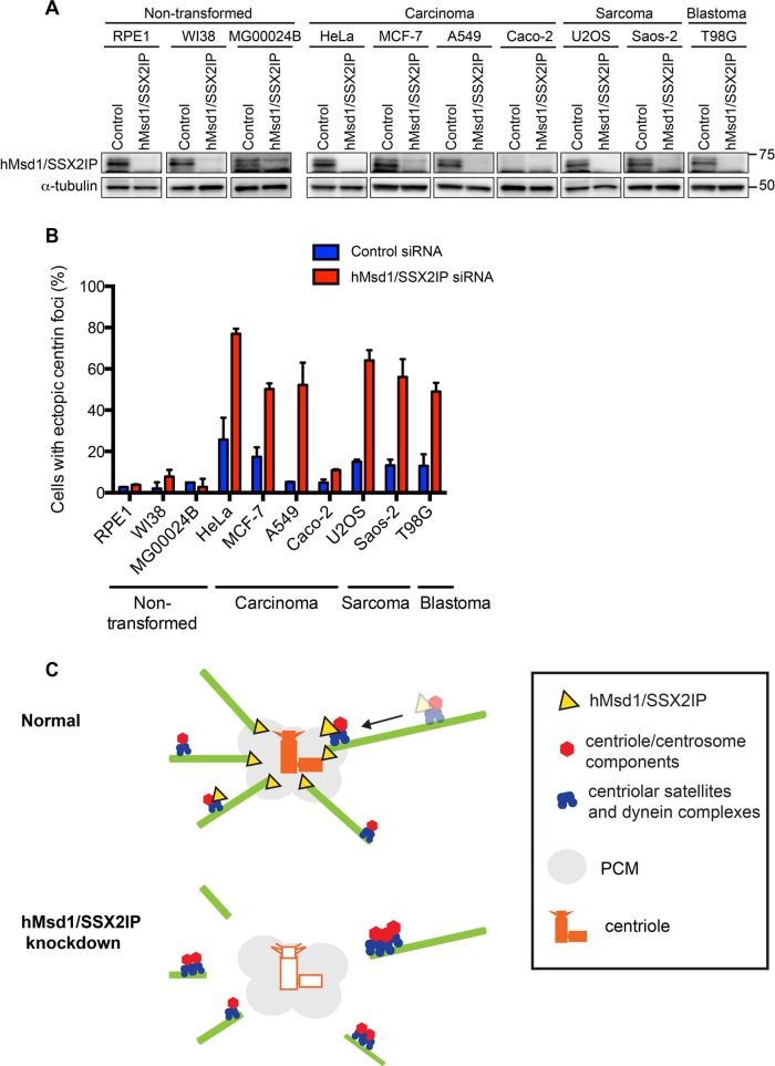
Centriolar satellites are numerous electron-dense granules dispersed around the centrosome. Mutations in their components are linked to various human diseases, but their molecular roles remain elusive. In particular, the significance of spatial communication between centriolar satellites and the centrosome is unknown. hMsd1/SSX2IP localizes to both the centrosome and centriolar satellites and is required for tethering microtubules to the centrosome. Here we show that hMsd1/SSX2IP-mediated microtubule anchoring is essential for proper centriole assembly and duplication. On hMsd1/SSX2IP knockdown, the centriolar satellites become stuck at the microtubule minus end near the centrosome. Intriguingly, these satellites contain many proteins that normally localize to the centrosome. Of importance, microtubule structures, albeit not being anchored properly, are still required for the emergence of abnormal satellites, as complete microtubule depolymerization results in the disappearance of these aggregates from the vicinity of the centrosome. We highlighted, using superresolution and electron microscopy, that under these conditions, centriole structures are faulty. Remarkably, these cells are insensitive to Plk4 overproduction-induced ectopic centriole formation, yet they accelerate centrosome reduplication upon hydroxyurea arrest. Finally, the appearance of satellite aggregates is cancer cell specific. Together our findings provide novel insights into the mechanism of centriole assembly and microtubule anchoring.


Figures

FIGURE 1:
FIGURE 1:
Microtubule anchoring of hMsd1/SSX2IP to the centrosome is essential for the scattered localization of PCM1. (A) Microtubule disorganization upon hMsd1/SSX2IP depletion leads to PCM1 aggregation. U2OS cells were transfected with control or hMsd1/SSX2IP siRNA and immunostained after 48 h with anti–α-tubulin (red) and anti-PCM1 antibodies (green). Regions outlined by squares in the top row are enlarged in the bottom two rows. DAPI staining (blue) is also shown in merged images (top). Scale bars, 10 μm, 5 μm. Schematics for PCM1 localization patterns are depicted below. (B, C) Quantification. The percentage of cells displaying PCM1 aggregation (B) and PCM1 signal intensities around the centrosome (C) were quantified. Twenty-five–pixel squares around the centrosome were measured. U2OS cells were treated with hMsd1/SSX2IP siRNA and simultaneously transfected with plasmids containing myc-PACT, myc-tagged full-length hMsd1/SSX2IP (hMsd1/SSX2IP-FL), or myc-PACT connected to the C-terminal half of hMsd1/SSX2IP (hMsd1/SSX2IP-C-PACT). Data represent mean ± SD (B, >300 cells derived from three independent experiments, n = 3; C, >100 cells, n = 3). Statistical analysis was performed using two-tailed unpaired Student's t tests. **p < 0.001, ***p < 0.0001; n.s., not significant. (D) PCM aggregates upon hMsd1/SSX2IP depletion stem from defects in microtubule anchoring of the centrosome. Cells were stained with antibodies against myc (blue), PCM1 (green), and α-tubulin (red). Enlarged images corresponding to regions marked with arrowheads (top) are shown at the bottom. Scale bars, 5 μm, 1 μm (enlarged images).
FIGURE 2:
FIGURE 2:
hMsd1/SSX2IP knockdown compromises dynamic motility of PCM1 and results in the aggregation that is dependent on microtubule structures. (A) PCM1 aggregates localize to the end of microtubules. Regions outlined by squares are enlarged, in which images of only one z-section are shown. Scale bars, 5 μm, 1 μm. (B) Microtubule depolymerization leads to the dispersed localization of PCM1 in hMsd1/SSX2IP-depleted cells. U2OS cells were transfected with control or hMsd1/SSX2IP siRNA and treated with DMSO (left) or nocodazole (right) for 2 h. Cells were then fixed and immunostained with antibodies against PCM1 (green) and α-tubulin (red). DNA was stained with DAPI (blue). Scale bars, 5 μm. (C) Quantification of cells displaying PCM1 aggregation around the centrosome. Data represent the mean ± SD (>150 cells, n = 3). ***p < 0.0001; n.s., not significant. (D) The trajectory of PCM1 signals. U2OS cells were treated with control or hMsd1/SSX2IP siRNA and further transfected with EGFP-PCM1 after 24 h. At 24 h after the second transfection, the cells were observed and time-lapse imaging performed (n = 17 in control siRNA cells; n = 16 in hMsd1/SSX2IP siRNA cells).
FIGURE 3:
FIGURE 3:
hMsd1/SSX2IP depletion leads to the formation of supernumerary centrin foci that are dependent on PCM1. (A) Extra centrin foci appear upon hMsd1/SSX2IP depletion in a PCM1-dependent manner. HeLa cells stably expressing centrin-GFP were cotransfected with control, hMsd1/SSX2IP, control and PCM1, or hMsd1/SSX2IP and PCM1 siRNAs. At 48 h after transfection, cells were fixed and immunostained with anti-GFP (green) and anti-PCM1 antibodies (red). DNA was stained with DAPI (blue). Scale bars, 5 μm, 1 μm (bottom squares). (B) Quantification of cells containing ectopic centrin foci. Data represent the mean ± SD (>300 cells, n = 3). ***p < 0.001; n.s., not significant. (C) Representative images of immuno-EM using an anti-GFP antibody in HeLa cells stably expressing centrin-GFP. Note that gold particles overlap with the electron-dense granules that represent centriolar satellites in hMsd1/SSX2IP-depleted cells (magenta arrows). Gold particles at the centriole (yellow arrowheads) in hMsd1/SSX2IP-depleted cells (n = 49) were fewer than with control cells (n = 61). Scale bar, 200 nm. (D) The centrosome-targeted C-terminal half of hMsd1/SSX2IP suppressed the formation of ectopic centrin foci. U2OS cells were cotransfected with hMsd1/SSX2IP siRNA and plasmids containing myc alone, siRNA-resistant, full-length myc-hMsd1/SSX2IP (myc-hMsd1/SSX2IP-FL), or myc-PACT connected C-terminal half of hMsd1/SSX2IP (myc-hMsd1/SSX2IP-C-PACT). Left, cells were stained with antibodies against myc (green) and centrin-2 (red). DNA was stained with DAPI (blue). Regions marked with arrowheads (top) are enlarged at the bottom. Scale bars, 5 μm, 1μm (bottom, enlarged images). Right, quantification of cells displaying ectopic centrin foci (>200 cells, n = 3). ***p < 0.0001, n.s., not significant.
FIGURE 4:
FIGURE 4:
A subset of centriolar/centrosomal components accumulates as aggregates around the centrosome and colocalize with PCM1. (A) Some centriolar/centrosomal components were amplified at the pericentriolar region. HeLa cells stably expressing centrin-GFP were transfected with control or hMsd1/SSX2IP siRNA, and 48 h after transfection, cells were fixed and immunostained with antibodies against individual proteins indicated (red) and centrin-GFP (green). (B) Quantification of cells containing ectopic foci of each centriolar/centrosomal component. Data represent the mean ± SD (>200 cells, n = 3). ***p < 0.0001, **p < 0.001. (C) Ectopic foci containing centrobin (left) or C-Nap1 (right) colocalize with PCM1 aggregates in hMsd1/SSX2IP-depleted cells. Cells were stained with antibodies against PCM1 (green) and centrobin or C-Nap1 (red). Scale bar, 1 μm.
FIGURE 5:
FIGURE 5:
Centriole structures are abnormal in hMsd1/SSX2IP-depleted cells. (A) Superresolution microscopy (OMX) images. Representative images show two normal ring structures of Cep152 in either control (top) or after hMsd1/SSX2IP depletion (bottom two rows), whereas centrobin displays abnormal non-ring structures in hMsd1/SSX2IP-depleted cells. In control cells, centrobin forms smaller rings juxtaposed with Cep152 rings (>20 cells, n = 2). Right, schematics depicting the localization patterns of centrobin and Cep152 together with predicted centriole structures (gray). Scale bar, 1 μm. (B) CLEM images. HeLa cells stably expressing centrin-GFP were transfected with control or hMsd1/SSX2IP siRNA and processed for CLEM analysis. Top row, correlative images of fluorescence (left) and EM (right). Bottom two rows, serial section images corresponding to the areas marked with yellow arrowheads in the top row (control siRNA n = 6, hMsd1/SSX2IP siRNA n = 12). Note that distinctive electron-dense centriole structures were more difficult to identify in the hMsd1/SSX2IP-depleted cells. Scale bars, 2 μm, 5 μm (top), 500 nm (bottom two rows). (C) Loss of hMsd1/SSX2IP suppresses the appearance of a rosette-like arrangement of extra centrins induced by myc-Plk4 overproduction. U2OS cells were transfected with control or hMsd1/SSX2IP siRNA, and plasmids containing myc-connected Plk4 constructs were further transfected after 24 h. Cells were stained with antibodies against myc (blue), centrin-2 (green), and γ-tubulin (red). Representative images of rosette formation in control siRNA cells (top) and two examples in hMsd1/SSX2IP siRNA cells (bottom two rows). One (middle) displays two to four centrin foci, whereas the other (bottom) exhibits the dispersed appearance of extra centrin foci, the phenotype reminiscent of hMsd1/SSX2IP depletion. Scale bar, 1 μm. (D) Quantification of cells containing a rosette-like arrangement of extra centrins. Data represent the mean ± SD (>200 cells, n = 2). ***p < 0.0001.
FIGURE 6:
FIGURE 6:
hMsd1/SSX2IP depletion promotes centrosome overduplication in HU-arrested U2OS cells. (A–C) HU was added to U2OS cells stably expressing centrin-GFP and simultaneously transfected with control or hMsd1/SSX2IP siRNA. At 48 and 72 h after transfection, cells were fixed and stained with antibodies against γ-tubulin (A), hSAS-6 (B), or Cep152 (C). (A, left) Images displaying three representative types of cells: ≤4 centrin/≤2 γ-tubulin (denoted as normal), >4 centrin/≤2 γ-tubulin (centrin amplification), and >4 centrin/>2 γ-tubulin (overduplication). Arrows mark centrin dots that colocalize with γ-tubulin. Scale bars, 5 μm. Quantification of the percentage of cells representing the three types of centrin/γ-tubulin (A, right), hSAS-6 (B), or Cep152 (C) patterns. Data represent the mean ± SD (>200 cells, n = 2). For immunofluorescence images corresponding to B and C, see Supplemental Figure S4, A and B, respectively. (D) HU was added to U2OS cells stably expressing centrin-GFP and simultaneously transfected with control or hMsd1/SSX2IP siRNA. At 48 h after transfection, cells were fixed and stained with antibodies against centrin-2 (blue), centrobin (green), and γ-tubulin (red). Left, the four representative types of cells: type I, ≤4 centrin, ≤4 centrobin, ≤2 γ-tubulin (normal); type II, >4 centrin, ≤4 centrobin, ≤2 γ-tubulin (centrin amplification), type III, >4 centrin, >4 centrobin, ≤2 γ-tubulin (centrin and centrobin amplification); and type IV, >4 centrin, >4 centrobin, >2 γ-tubulin (overduplication). Right, quantification of the percentage of cells representing the four types of centrin/centrobin/γ-tubulin patterns. Data represent the mean ± SD (>200 cells, n = 2). Note that in type III, most, if not all, amplified centrin foci overlapped with those of centrobin, whereas in type IV, this is also the case for γ-tubulin extra foci.
FIGURE 7:
FIGURE 7:
Centrin amplification upon hMsd1/SSX2IP knockdown is cancer cell specific. (A) Nontransformed (RPE1, W138, MG00024B), carcinoma (HeLa, MCF-7, A549, Caco-2), sarcoma (U2OS, Saos-2), or blastoma cell lines (T98G) were treated with control or hMsd1/SSX2IP siRNA. Immunoblotting with an anti-hMsd1/SSX2IP antibody is shown for each cell line with α-tubulin used as a loading control. (B) Quantification of cells containing ectopic centrin foci. Data represent the mean ± SD (>150 cells, n = 2). (C) A scheme depicting the transport and accumulation of centriolar components in control and hMsd1/SSX2IP-depleted cells. Top, under normal conditions, hMsd1/SSX2IP (yellow) localizes to centriolar satellites (blue) and around the centrosome (gray area), and is responsible for anchoring the minus end of microtubules to the centrosome. Centriolar satellites transport a cohort of centriolar/centrosomal components (red) toward the centrosome via microtubules. This process is essential to the assembly of intact centriole structures (orange). Bottom, in the absence of hMsd1/SSX2IP, particularly in cancer cells, microtubules are no longer tethered to the centrosome. Consequently, centriolar satellites carrying centriolar/centrosomal components become stuck at the end of the microtubules, leading to faulty assembly of centrioles (outlined in orange). Note that our work indicates that in the absence of hMsd1/SSX2IP-mediated microtubule anchoring, particularly in cancer cells, other independent pathways responsible for centriole assembly, such as diffusion-based protein–protein interaction, are not sufficient to build centrioles properly. At the moment, however, we cannot explicitly conclude whether the sole reason for faulty centriole assembly is attributed to the defects in the release of the centriolar/centrosomal proteins from centriolar satellites. By contrast, in nontransformed cells, microtubule disorganization is observed; however, the role of hMsd1/SSX2IP in centriole assembly is cryptic, and alternate compensatory pathways may be exploited in these cells.

Similar articles

Cited by

References

    1. Azimzadeh J, Marshall WF. Building the centriole. Curr Biol. 2010;20:R816–R825. - PMC - PubMed
    1. Balczon R, Bao L, Zimmer WE. PCM-1, A 228-kD centrosome autoantigen with a distinct cell cycle distribution. J Cell Biol. 1994;124:783–793. - PMC - PubMed
    1. Balczon R, Bao L, Zimmer WE, Brown K, Zinkowski RP, Brinkley BR. Dissociation of centrosome replication events from cycles of DNA synthesis and mitotic division in hydroxyurea-arrested Chinese hamster ovary cells. J Cell Biol. 1995;130:105–115. - PMC - PubMed
    1. Balestra FR, Strnad P, Fluckiger I, Gonczy P. Discovering regulators of centriole biogenesis through siRNA-based functional genomics in human cells. Dev Cell. 2013;25:555–571. - PubMed
    1. Barenz F, Inoue D, Yokoyama H, Tegha-Dunghu J, Freiss S, Draeger S, Mayilo D, Cado I, Merker S, Klinger M, et al. The centriolar satellite protein SSX2IP promotes centrosome maturation. J Cell Biol. 2013;202:81–95. - PMC - PubMed

Publication types

MeSH terms

LinkOut - more resources