Review
. 2015 Apr 1;7(4):a017970.
doi: 10.1101/cshperspect.a017970.

Meiosis and maternal aging: insights from aneuploid oocytes and trisomy births

Affiliations
Review

Meiosis and maternal aging: insights from aneuploid oocytes and trisomy births

Mary Herbert et al. Cold Spring Harb Perspect Biol. .

Abstract

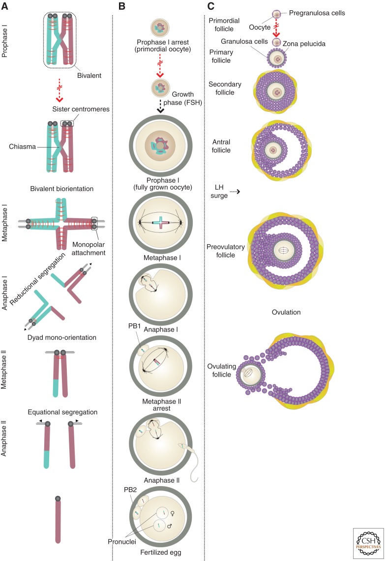
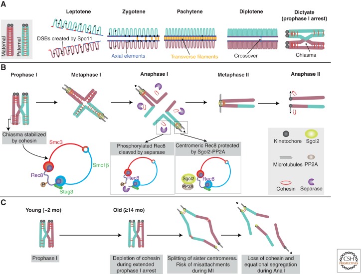
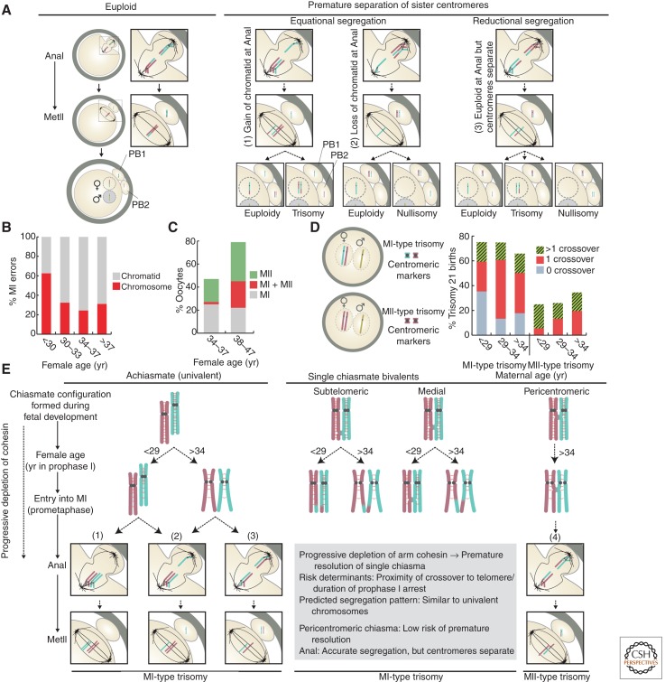
In most organisms, genome haploidization requires reciprocal DNA exchanges (crossovers) between replicated parental homologs to form bivalent chromosomes. These are resolved to their four constituent chromatids during two meiotic divisions. In female mammals, bivalents are formed during fetal life and remain intact until shortly before ovulation. Extending this period beyond ∼35 years greatly increases the risk of aneuploidy in human oocytes, resulting in a dramatic increase in infertility, miscarriage, and birth defects, most notably trisomy 21. Bivalent chromosomes are stabilized by cohesion between sister chromatids, which is mediated by the cohesin complex. In mouse oocytes, cohesin becomes depleted from chromosomes during female aging. Consistent with this, premature loss of centromeric cohesion is a major source of aneuploidy in oocytes from older women. Here, we propose a mechanistic framework to reconcile data from genetic studies on human trisomy and oocytes with recent advances in our understanding of the molecular mechanisms of chromosome segregation during meiosis in model organisms.


Figures

Figure 1.
Figure 1.
Meiotic divisions and mammalian oogenesis. (A) Bivalent chromosome consisting of replicated maternal and paternal homologs linked by a crossover (chiasma). Accurate segregation of homologs during meiosis I (MI) depends on biorientation of bivalents, which necessitates monopolar attachment of sister kinetochores. Dissolution of cohesion from chromosome arms during anaphase I (anaI) converts bivalents to dyads. Dissolution of centromeric cohesion results in segregation of chromatids during anaphase II. (B,C) Following the formation of bivalent chromosomes, oocytes arrest in prophase of MI surrounded by a layer of pregranulosa cells to form primordial follicles. Primordial follicles are recruited for growth on an ongoing basis. Growing follicles do not develop to the preovulatory stage until after puberty when they become responsive to follicle-stimulating hormone (FSH). Fully grown oocytes enter M phase following a surge of luteinizing hormone (LH). During anaphase I, the outermost dyads are ejected in the first polar body (PB1). The dyads remaining in the oocyte realign on the meiosis II (MII) spindle. The oocyte is then ovulated and remains arrested at metaphase of MII until sperm entry triggers anaphase II when half of the chromatids are ejected in the second polar body (PB2). Fertilization is marked by formation of pronuclei, in which the maternal and paternal haploid genomes are separately packaged.
Figure 2.
Figure 2.
Molecular regulation of meiotic chromosome structure. (A) Replicated maternal and paternal homologs undergo meiotic recombination following entry into prophase I. The process is initiated by Spo11-mediated double-strand break (DSB) formation. Axial elements support DSB formation and DNA repair from the homolog. The synaptonemal complex (SC) promotes formation of double Holliday junctions (dHJs), which are resolved to crossovers. Crossovers become visible as chiasmata in diplotene and oocytes then enter dictyate (prophase I arrest). (B) Bivalent chromosomes are stabilized by arm cohesin distal to the chiasma. The cohesin ring shown contains meiosis-specific subunits: the α-kleisin Rec8, Smc1β, forms a heterodimer with Smc3 and Stag3, which regulates stability of Rec8-containing complexes. During anaphase I, separase cleaves phosphorylated Rec8 on the chromosome arms. Cohesin at the centromere is protected by the phosphatase PP2A, which is recruited there by Sgo12. Dyad chromosomes align on the meiosis II (MII) spindle and cohesin at the centromere is cleaved during anaphase II. (C) Schematic summarizing the main elements of the “cohesin deterioration hypothesis,” based on findings in mouse oocytes. Cohesin loaded in early oogenesis becomes depleted during prolonged arrest in prophase I. This results in loss of bivalent structure, which, in mice, manifests as distally associated homologs. Sister centromeres also lose their unified structure and frequently undergo premature resolution followed by equational (shown) or reductional segregation.
Figure 3.
Figure 3.
Chromosome structure and missegregation in human oocytes and maternal trisomy. (A) Schematic showing the types of aneuploidy involving premature separation of sister centromeres described in human oocytes. (B) Graph showing the proportions of whole chromosome and chromatid errors reported from cytogenetic analysis of meiosis II (MII)-arrested human oocytes (n = 1397) from women of different ages. (Calculated from data in Pellestor et al. 2003.) (C) Graph showing the incidence of segregation errors occurring during each of the meiotic divisions in oocytes from younger and older women thought to be at high risk of meiotic aneuploidy. Based on array-comparative genome hybridization (CGH) analysis of both polar bodies from human oocytes/zygotes (n = 420) (Fragouli et al. 2013). (D) Schematic showing meiosis I (MI)- and meiosis II (MII)-type trisomy based on pericentromeric markers in the two maternally inherited copies. Graph shows the number of crossovers observed in MI- and MII-type trisomy 21 children across three maternal age groups (Oliver et al. 2008). (E) Model showing the predicted effect of a progressive depletion of arm cohesion on the structure of univalents and single chiasmate bivalents. Depletion of arm cohesion distal to the chiasma is predicted to induce its premature resolution. The threshold level of cohesin required to maintain bivalent stability depends on the distance between the chiasma and the telomere. Bivalents with pericentromeric chiasma are, therefore, predicted to be more stable than those with a more distally posited chiasma. The predicted segregation patterns during MI are based on (1) evidence that premature separation of sister centromeres is the major cause of aneuploidy in human oocytes, (2) data from genetic analyses of trisomy 21 showing the incidence of MI- and MII-type errors associated with each of the chiasmate configurations stratified by maternal age (Oliver et al. 2008), and (3) findings in yeast meiosis (Sakuno et al. 2011) and mouse oocytes (Kouznetsova et al. 2007) that sister kinetochores of univalent chromosomes establish bipolar attachments on the MI spindle.

References

    1. Adhikari D, Liu K 2009. Molecular mechanisms underlying the activation of mammalian primordial follicles. Endocr Rev 30: 438–464. - PubMed
    1. Angell R 1991. Predivision in human oocytes at meiosis I: A mechanism for trisomy formation in man. Hum Genet 86: 383–387. - PubMed
    1. Angell R 1997. First meiotic division non-disjunction in human oocytes. Am J Hum Genet 61: 23–32. - PMC - PubMed
    1. Azoury J, Lee KW, Georget V, Rassinier P, Leader B, Verlhac MH 2008. Spindle positioning in mouse oocytes relies on a dynamic meshwork of actin filaments. Curr Biol 18: 1514–1519. - PubMed
    1. Baart EB, Martini E, Eijkemans MJ, Van Opstal D, Beckers NGM, Verhoeff A, Macklon NS, Fauser BCJM 2007. Milder ovarian stimulation for in vitro fertilization reduces aneuploidy in the human preimplantation embryo: A randomized controlled trial. Hum Reprod 22: 980–988. - PubMed

Publication types