Review
. 2015 Apr 1;5(4):a021352.
doi: 10.1101/cshperspect.a021352.

Animal models and the molecular biology of hepadnavirus infection

Affiliations
Review

Animal models and the molecular biology of hepadnavirus infection

William S Mason. Cold Spring Harb Perspect Med. .

Abstract

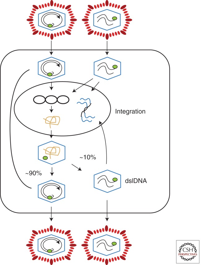
Australian antigen, the envelope protein of hepatitis B virus (HBV), was discovered in 1967 as a prevalent serum antigen in hepatitis B patients. Early electron microscopy (EM) studies showed that this antigen was present in 22-nm particles in patient sera, which were believed to be incomplete virus. Complete virus, much less abundant than the 22-nm particles, was finally visualized in 1970. HBV was soon found to infect chimpanzees, gorillas, orangutans, gibbon apes, and, more recently, tree shrews (Tupaia belangeri) and cynomolgus macaques (Macaca fascicularis). This restricted host range placed limits on the kinds of studies that might be performed to better understand the biology and molecular biology of HBV and to develop antiviral therapies to treat chronic infections. About 10 years after the discovery of HBV, this problem was bypassed with the discovery of viruses related to HBV in woodchucks, ground squirrels, and ducks. Although unlikely animal models, their use revealed the key steps in hepadnavirus replication and in the host response to infection, including the fact that the viral nuclear episome is the ultimate target for immune clearance of transient infections and antiviral therapy of chronic infections. Studies with these and other animal models have also suggested interesting clues into the link between chronic HBV infection and hepatocellular carcinoma.


Figures

Figure 1.
Figure 1.
The genome and endogenous polymerase reaction of hepatitis B virus (HBV). (A) The HBV genome is a partially double-stranded DNA, held in a relaxed circular conformation by a short cohesive overlap between the 5′ ends of the two DNA strands (Summers et al. 1975). One strand, later found to be the plus strand, is always incomplete in virus particles, with a gap that may encompass up to 50% of the genome length. The minus strand is always complete. The large circle at the 5′ end of the minus strand represents a covalently attached protein (Gerlich and Robinson 1980) that was later shown to be the viral DNA polymerase/reverse transcriptase (Bartenschlager and Schaller 1988). Pregenomic RNA, the template for viral DNA synthesis, is shown for comparison. DR1 and DR2 are 11-nucleotide direct repeats (12-nucleotide for duck hepatitis B virus [DHBV]) on the pregenome that play essential roles in priming of viral DNA synthesis (see text). (B) HBV and other hepadnaviruses contain the viral DNA polymerase, which can fill in the single-stranded gap in vitro (Summers et al. 1975). The fill-in reaction can be performed by pelleting virus from serum, adding nonionic detergent and radiolabeled deoxynucleotides (dNTPs) to the pellet, and incubating at 37°C. The radiolabeled DNA can be detected by agarose gel electrophoresis and autoradiography, as shown here for DHBV and woodchuck hepatitis virus (WHV). This assay was instrumental in the discovery of WHV, DHBV, and ground squirrel hepatitis virus (GSHV) (Summers et al. 1978; Marion et al. 1980; Mason et al. 1980).
Figure 2.
Figure 2.
Hepadnavirus phylogeny. (A) The orthohepadnaviruses infect mammals and share similarities in genome sequence and open reading frame location, size, and function. Four species have been defined, with hepatitis B virus (HBV), woolly monkey HBV (WMHBV), ground squirrel hepatitis virus (GSHV), and woodchuck hepatitis virus (WHV) serving as the prototype for each species. As shown, there are at least eight different genotypes of human HBV and additional genotypes in chimpanzees, orangutans, and gibbon apes. WMHBV, WHV, and GSHV are designated as distinct species based on differences in sequence and a unique host range. The arctic squirrel hepatitis virus (ASHV) is most similar to GSHV, but it is not yet known whether it has a host range distinct from that of GSHV or WHV. (B) The Avihepadnavirus isolates to date come mostly from ducks and geese. Viruses that are not from domestic ducks but considered to be of the same species as duck HBV (DHBV) and Chi-tung County (CC)-DHBV isolates from China include isolates from the snow goose (SGHBV), Orinoco sheldgoose (OSHBV), ashy-headed sheldgoose (ASHBV), Puna teal (PTHBV), and Chiloé wigeon (CWHBV) (Guo et al. 2005). DHBV is also found in wild mallards (Cova et al. 1986), from which most domesticated ducks, except Muscovy, were derived. Of the remaining avihepadnaviruses shown here, only heron HBV (HHBV) (Sprengel et al. 1988) is designated a distinct species (Fauquet et al. 2005). Virus isolates from the Ross goose (RGHBV), crane (CHBV) (Prassolov et al. 2003), stork (STHBV) (Pult et al. 2001), and parakeet (PHBV) (Piasecki et al. 2013) remain unassigned.
Figure 3.
Figure 3.
Comparison of retrovirus and hepadnavirus DNA synthesis pathways. (A) Moloney murine leukemia virus (MoMLV) DNA synthesis. Reverse transcription of MoMLV RNA begins when a cell is infected. The primer for reverse transcription is the 3′ hydroxyl (OH) of tRNAPro, which is annealed near to the 5′ end of the viral RNA genome via hybridization to an 18-nucleotide primer binding site (PBS) (a). Synthesis extends to the 5′ end of the viral RNA. The reverse-transcribed RNA sequences are degraded by the viral RNase H (b,c). At this point, the DNA product can hybridize to the 3′ end of viral RNA through the 68-nucleotide R domain, found at both ends of viral RNA (d). This facilitates reverse transcription of the remainder of the RNA template. Plus-strand synthesis initiates near the 5′ end of the minus strand from an RNA oligonucleotide that is left behind during RNase H degradation of the reverse-transcribed viral RNA (e). (It should be noted that some retroviruses prime plus-strand synthesis from multiple sites, including the polypurine tract [PPT] region as well as upstream sites.) Plus-strand synthesis extends rightward to copy the 3′ 18 nucleotides of the tRNA, recreating the PBS as DNA. Once minus-strand synthesis has made a complementary copy of the PBS (f), the 3′ ends of the nascent plus and minus strands can hybridize to form a circle (g). Minus-strand synthesis extends to the 5′ end of the plus strand, presumably via strand displacement synthesis (h), and plus-strand synthesis extends to the 5′ end of the minus strand to create a DNA with a large terminal redundancy (LTR; U3-R-U5). This terminally redundant linear DNA is the substrate for integration into host DNA. Integrated DNA serves as the provirus template for new viral RNA synthesis. (B) duck hepatitis B virus (DHBV) DNA synthesis. In this early model (Lien et al. 1986), reverse transcription is primed from a tyrosine residue of a protein primer, later shown to map to the terminal protein domain of the viral DNA polymerase (a), and begins within the 3′ copy of DR1. Minus-strand synthesis extends to the 5′ end of the pregenome (b,c). Because it began in R and extended almost to the 5′ end of the RNA template, the nascent minus strand is terminally redundant by 7–8 nucleotides. The pregenome is degraded by RNase H, leaving an 18-nucleotide sequence including the 5′ CAP and first 18 nucleotides of the pregenome (c), including the 12-base repeat, DR1, which is present in the terminal redundancy of the pregenome. This oligoribonucleotide is then translocated from DR1 to DR2, which maps upstream of the 3′ terminal redundancy (d). The oligoribonucleotide hybridizes to DR2 on the minus-strand DNA because DR1 and DR2 are identical in sequence. Plus-strand synthesis initiates at the 3′ end of DR2 and extends to the end of the minus strand. Circularization to continue plus-strand synthesis is apparently facilitated by the 7- to 8-nucleotide terminal redundancy on the minus strand (e,f). Synthesis continues, to produce a partially double-stranded virus genome (with DHBV, most plus strands are nearly full-length, excluding DR2, to which the plus-strand primer remains bound). Approximately 10% of the time, the plus-strand primer is not translocated to DR2, and plus-strand synthesis begins at the upstream copy of DR1, resulting in a linear virus genome (see Fig. 4).
Figure 3.
Figure 3.
Comparison of retrovirus and hepadnavirus DNA synthesis pathways. (A) Moloney murine leukemia virus (MoMLV) DNA synthesis. Reverse transcription of MoMLV RNA begins when a cell is infected. The primer for reverse transcription is the 3′ hydroxyl (OH) of tRNAPro, which is annealed near to the 5′ end of the viral RNA genome via hybridization to an 18-nucleotide primer binding site (PBS) (a). Synthesis extends to the 5′ end of the viral RNA. The reverse-transcribed RNA sequences are degraded by the viral RNase H (b,c). At this point, the DNA product can hybridize to the 3′ end of viral RNA through the 68-nucleotide R domain, found at both ends of viral RNA (d). This facilitates reverse transcription of the remainder of the RNA template. Plus-strand synthesis initiates near the 5′ end of the minus strand from an RNA oligonucleotide that is left behind during RNase H degradation of the reverse-transcribed viral RNA (e). (It should be noted that some retroviruses prime plus-strand synthesis from multiple sites, including the polypurine tract [PPT] region as well as upstream sites.) Plus-strand synthesis extends rightward to copy the 3′ 18 nucleotides of the tRNA, recreating the PBS as DNA. Once minus-strand synthesis has made a complementary copy of the PBS (f), the 3′ ends of the nascent plus and minus strands can hybridize to form a circle (g). Minus-strand synthesis extends to the 5′ end of the plus strand, presumably via strand displacement synthesis (h), and plus-strand synthesis extends to the 5′ end of the minus strand to create a DNA with a large terminal redundancy (LTR; U3-R-U5). This terminally redundant linear DNA is the substrate for integration into host DNA. Integrated DNA serves as the provirus template for new viral RNA synthesis. (B) duck hepatitis B virus (DHBV) DNA synthesis. In this early model (Lien et al. 1986), reverse transcription is primed from a tyrosine residue of a protein primer, later shown to map to the terminal protein domain of the viral DNA polymerase (a), and begins within the 3′ copy of DR1. Minus-strand synthesis extends to the 5′ end of the pregenome (b,c). Because it began in R and extended almost to the 5′ end of the RNA template, the nascent minus strand is terminally redundant by 7–8 nucleotides. The pregenome is degraded by RNase H, leaving an 18-nucleotide sequence including the 5′ CAP and first 18 nucleotides of the pregenome (c), including the 12-base repeat, DR1, which is present in the terminal redundancy of the pregenome. This oligoribonucleotide is then translocated from DR1 to DR2, which maps upstream of the 3′ terminal redundancy (d). The oligoribonucleotide hybridizes to DR2 on the minus-strand DNA because DR1 and DR2 are identical in sequence. Plus-strand synthesis initiates at the 3′ end of DR2 and extends to the end of the minus strand. Circularization to continue plus-strand synthesis is apparently facilitated by the 7- to 8-nucleotide terminal redundancy on the minus strand (e,f). Synthesis continues, to produce a partially double-stranded virus genome (with DHBV, most plus strands are nearly full-length, excluding DR2, to which the plus-strand primer remains bound). Approximately 10% of the time, the plus-strand primer is not translocated to DR2, and plus-strand synthesis begins at the upstream copy of DR1, resulting in a linear virus genome (see Fig. 4).
Figure 4.
Figure 4.
Hepadnavirus infection. Virus with a relaxed circular (RC) genome is shown on the top right. Upon infection, the DNA is translocated to the nucleus, where the RC DNA genome is converted to covalently closed circular DNA (cccDNA), the template for viral RNA synthesis. When one of the largest viral RNAs, the pregenome, enters the cytoplasm, it can be packaged into viral nucleocapsids along with the viral reverse transcriptase and copied to produce new RC DNA. In the first few days of infection, newly made DNA is transported to the nucleus to amplify cccDNA copy number. As envelope proteins accumulate, this pathway is shut down and the nucleocapsids with RC DNA are enveloped and exported from the cell (Summers et al. 1990; Lenhoff and Summers 1994). Virus with linear DNA (Fig. 3) is also infectious and can form cccDNA via nonhomologous recombination, leading to defective cccDNAs. Linear genomes are also the substrate for integration of viral DNA into host DNA (Gong et al. 1995, 1999; Yang and Summers 1995, 1999). Integration is random on the host genome but appears to occur preferentially near the ends of linear viral DNAs. Some integrants also appear derived from RC DNA that has been linearized by displacement synthesis of plus and minus strands through the cohesive overlap, to create a linear DNA with a large terminal redundancy (Yang et al. 1996). dslDNA, double-stranded linear DNA.

References

    1. Aden DP, Fogel A, Plotkin S, Damjanov I, Knowles BB 1979. Controlled synthesis of HBsAg in a differentiated human liver carcinoma-derived cell line. Nature 282: 615–616. - PubMed
    1. Alison MR, Islam S, Lim S 2009. Stem cells in liver regeneration, fibrosis and cancer: The good, the bad and the ugly. J Pathol 217: 282–298. - PubMed
    1. Bartenschlager R, Schaller H 1988. The amino-terminal domain of the hepadnaviral P-gene encodes the terminal protein (genome-linked protein) believed to prime reverse transcription. EMBO J 7: 4185–4192. - PMC - PubMed
    1. Bayer ME, Blumberg BS, Werner B 1968. Particles associated with Australia antigen in the sera of patients with leukaemia, Down’s syndrome and hepatitis. Nature 218: 1057–1059. - PubMed
    1. Beasley RP, Lin CC, Hwang LY, Chien CS 1981. Hepatocellular carcinoma and hepatitis B virus. Lancet 2: 1129–1133. - PubMed