. 2015 May 22;290(21):13417-26.
doi: 10.1074/jbc.M114.633883. Epub 2015 Apr 1.

The N-terminal Set-β Protein Isoform Induces Neuronal Death

Affiliations

The N-terminal Set-β Protein Isoform Induces Neuronal Death

Ephraim F Trakhtenberg et al. J Biol Chem. .

Abstract

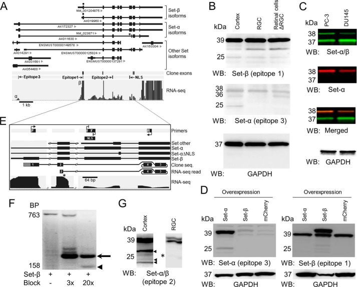
Set-β protein plays different roles in neurons, but the diversity of Set-β neuronal isoforms and their functions have not been characterized. The expression and subcellular localization of Set-β are altered in Alzheimer disease, cleavage of Set-β leads to neuronal death after stroke, and the full-length Set-β regulates retinal ganglion cell (RGC) and hippocampal neuron axon growth and regeneration in a subcellular localization-dependent manner. Here we used various biochemical approaches to investigate Set-β isoforms and their role in the CNS, using the same type of neurons, RGCs, across studies. We found multiple alternatively spliced isoforms expressed from the Set locus in purified RGCs. Set transcripts containing the Set-β-specific exon were the most highly expressed isoforms. We also identified a novel, alternatively spliced Set-β transcript lacking the nuclear localization signal and demonstrated that the full-length (∼39-kDa) Set-β is localized predominantly in the nucleus, whereas a shorter (∼25-kDa) Set-β isoform is localized predominantly in the cytoplasm. Finally, we show that an N-terminal Set-β cleavage product can induce neuronal death.

Keywords: Set-β; alternative splicing; cell death; neuron; protein translocation; subcellular fractionation.


Figures

FIGURE 1.
FIGURE 1.
Expression of Set-α and Set-β isoforms in primary neurons and a novel alternatively spliced Set-β isoform lacking the NLS. A, alternatively spliced Set isoforms from public databases aligned to the genome. Identification is indicated under the transcripts. Epitopes for Set antibodies used through the study are indicated. Epitope 1 is unique to Set-β-specific exon (), epitope 3 is unique to the Set-α specific exon, and epitope 2 is found in both isoforms. The asterisk indicates a Set-α isoform lacking an NLS sequence (Set-αΔNLS). Poly(A)-selected RNA-seq reads from purified P5 RGCs mapped uniquely to the Set locus (the raw data reads used for construction of this figure are available upon request). Reads aligned to the Set-β-specific exon (β) showed a significantly higher peak compared with the Set-α-specific exon (α). Transcripts and alignment to the genome were visualized using the Genomatix genome browser. Set locus RNA-seq raw reads data are available upon request. B, immunoblotting of P5 cortical homogenates, purified RGCs, and retinal cell-ΔRGC (as marked) for Set-β (epitope 1) and Set-α (epitope 3) revealed the predominance of full-length Set-β isoforms at 39 kDa and a weaker 25-kDa signal across samples. Set-α (epitope 3) detected lower molecular weight bands in cortical homogenates and retinal cell-ΔRGC but not in RGCs. WB, Western blot. C, human cell-lines, PC-3 and DU145, probed with anti-Set-α (epitope 3) and anti-Set-α/β antibodies, demonstrate the specificity of anti-Set-α antibody, which identified only one of two isoforms detected by the nonspecific anti-Set-α/β antibody. D, HEK293 cell line transfected with Set-α, Set-β, or mCherry constructs, probed with anti-Set-α (epitope 3) and anti-Set-β (epitope 1) antibodies, demonstrate the specificity of, and lack of cross-reactivity between, anti-Set-α and anti-Set-β antibodies. E, schematic of RT-PCR using a Set-β-specific forward primer (F), a reverse primer (R) from the exon past the NLS, and internally nested forward and reverse blocking primers against the NLS sequence modified at their 3′ ends to prevent PCR product elongation (f and r). RNA-seq read is 100% matching sequence identified in RNA-seq raw reads data. The lined area at 3′ corresponds to the 8-bp sequence past the clone sequence, which also aligned 100% to the exon. F, RT-PCR on RNA from acutely purified P5 RGCs showed increasing detection of a 158-bp product (arrowhead, clone seq in B) over the 763-bp band corresponding to the full-length Set-β at increasing concentrations of blocking primers 3 times and 20 times more than the Set-β primers. A higher molecular weight band (arrow) resulted from transcription initiated by the blocking primers (confirmed by sequencing because of impurities or imperfect blockade). G, immunoblotting of P5 cortical homogenates and purified RGCs (as marked) for Set-α/β (epitope 2) indicated the predominance of 39-kDa band, consistent with the full-length Set-β isoforms, and a weaker 25-kDa signal (asterisk) in both samples. Additional lower molecular weight bands (arrowheads) were detected in cortical homogenates but not in RGCs.
FIGURE 2.
FIGURE 2.
Set-β expression and developmental regulation in RGCs. A, P8 retinal sections were immunostained for Set-β (epitope 1), Set-α/β (epitope 2), and Brn3A (RGC marker), and counterstained with DAPI (nuclear marker) as marked. Example RGCs and their nuclei are outlined with dashed white lines in enlarged images. Scale bar = 100 μm (main panels) and 20 μm (enlarged images). GCL, ganglion cell layer; INL, inner nuclear layer. B, analysis of nuclear and cytoplasmic average pixel intensity of Set epitope 1 and 2 immunofluorescence in RGCs showed that Set-β (epitope 1) immunoreactivity is predominantly nuclear, whereas Set-α/β (epitope 2) immunoreactivity is predominantly cytoplasmic (n = 3, ≥85 randomly selected RGCs/experiment. Data are mean ± S.E. *, p < 0.01 by two-tailed Student's. C, P8 RGCs immunostained after 1 day in culture for endogenous Set-β (epitope 1) and Set-α/β (epitope 2) (green), Tuj1 (neuronal marker, red), and DAPI (nuclear marker). Scale bar = 20 μm. Nuclei are outlined with dashed white lines.
FIGURE 3.
FIGURE 3.
Set-β protein isoforms demonstrate differential localization. A–C, P5 RGC nuclear (N) and cytoplasmic (C) fractions immunoblotted for GAPDH (cytoplasmic marker), histone H3 (nuclear marker) (A), and Set-β (epitope 1) (B) and Set-α/β (epitope 2) (C) revealed that the full-length 39-kDa Set-β is predominantly nuclear, whereas the 25-kDa Set-β isoform is predominantly cytoplasmic in acutely purified RGCs. WB, Western blot.
FIGURE 4.
FIGURE 4.
Set-βΔC localizes to the cytoplasm and initiates cell death in cultured RGCs. A, full-length and truncated Set-β constructs, highlighting the serine 9 phosphorylation site (P), β isoform-specific antibody epitope (Y), PP2A inhibitory domain, NLS containing lysine 176 cleavage site (scissors), and acidic C-terminal domain deleted in the mutant construct (Set-βΔC). B, acutely purified P4 RGCs transfected with mCherry, full-length Set-β, or Set-βΔC were immunostained at 1, 2, and 3 days for fusion reporter tags (red) and Tuj1 (neurite marker, green) and counterstained with DAPI (nuclear marker, blue). After 1 day in culture, full-length Set-β localized to the nucleus, whereas Set-βΔC localized to both the nucleus and cytoplasm. By 2–3 days, morphologic characteristics of cell death, including cytoplasmic swelling (arrow) and cellular fragmentation (arrowhead), were apparent in Set-βΔC-transfected RGCs. Nuclei are outlined with white dashed lines. Scale bar = 20 μm. C, RGC survival after transfection was assessed by counting calcein-positive (live cell marker) or sytox-positive (dead cell marker) RGCs counterstained with Hoechst (nuclear marker). Example pictures from 2 h are shown. D, 2 h after transfection with full-length Set-β, Set-βΔC, or mCherry, survival was similar across conditions (n = 3, ≥1300 cells/condition in each experiment). Data are mean ± S.E. N.S., not significant by analysis of variance. E, at 1, 2, and 3 days, transfected RGCs were counted per unit area normalized to 1 day (106 cells/condition). Set-βΔC significantly increased cell death. Data are mean ± S.E. p < 0.01 by analysis of variance with repeated measures with post hoc LSD.

References

    1. Switzer C. H., Cheng R. Y., Vitek T. M., Christensen D. J., Wink D. A., Vitek M. P. (2011) Targeting SET/I(2)PP2A oncoprotein functions as a multi-pathway strategy for cancer therapy. Oncogene 30, 2504–2513 - PMC - PubMed
    1. von Lindern M., van Baal S., Wiegant J., Raap A., Hagemeijer A., Grosveld G. (1992) Can, a putative oncogene associated with myeloid leukemogenesis, may be activated by fusion of its 3′ half to different genes: characterization of the set gene. Mol. Cell. Biol. 12, 3346–3355 - PMC - PubMed
    1. Seo S. B., McNamara P., Heo S., Turner A., Lane W. S., Chakravarti D. (2001) Regulation of histone acetylation and transcription by INHAT, a human cellular complex containing the set oncoprotein. Cell 104, 119–130 - PubMed
    1. Gamble M. J., Fisher R. P. (2007) SET and PARP1 remove DEK from chromatin to permit access by the transcription machinery. Nat. Struct. Mol. Biol. 14, 548–555 - PubMed
    1. De Koning L., Corpet A., Haber J. E., Almouzni G. (2007) Histone chaperones: an escort network regulating histone traffic. Nat. Struct. Mol. Biol. 14, 997–1007 - PubMed

Publication types

MeSH terms

LinkOut - more resources