. 2015 May 15;290(20):12585-94.
doi: 10.1074/jbc.M114.625673. Epub 2015 Apr 1.

EDD, a ubiquitin-protein ligase of the N-end rule pathway, associates with spindle assembly checkpoint components and regulates the mitotic response to nocodazole

Affiliations

EDD, a ubiquitin-protein ligase of the N-end rule pathway, associates with spindle assembly checkpoint components and regulates the mitotic response to nocodazole

Flavia Scialpi et al. J Biol Chem. .

Abstract

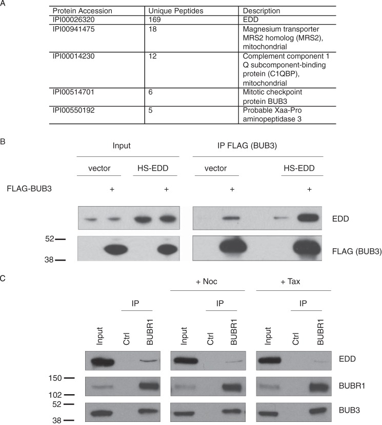
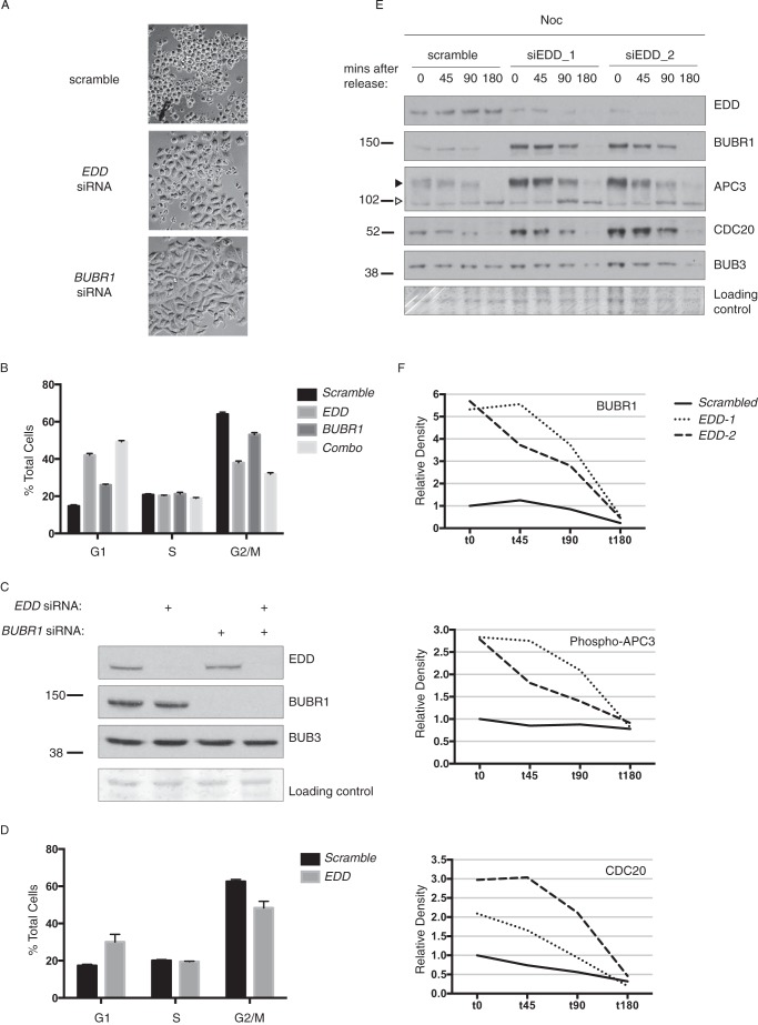
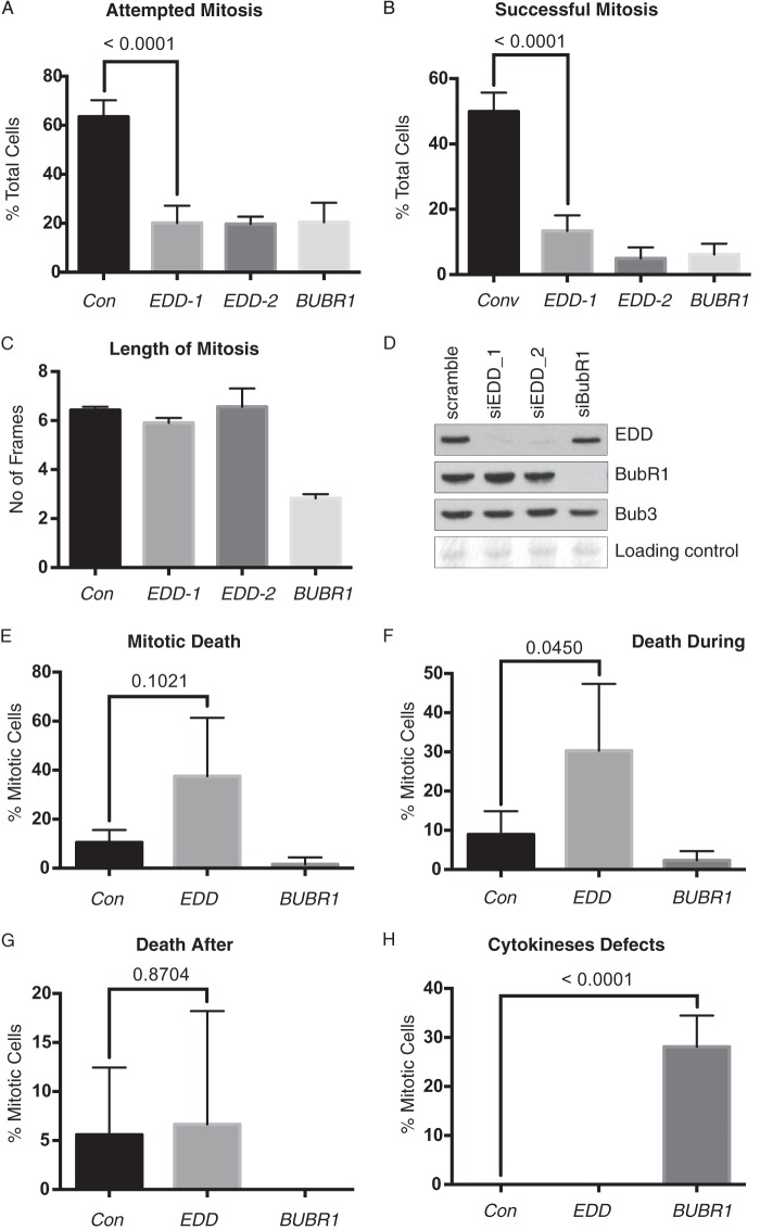
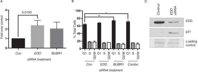
In this work, we identify physical and genetic interactions that implicate E3 identified by differential display (EDD) in promoting spindle assembly checkpoint (SAC) function. During mitosis, the SAC initiates a mitotic checkpoint in response to chromosomes with kinetochores unattached to spindle pole microtubules. Similar to Budding uninhibited by benzimidazoles-related 1 (BUBR1) siRNA, a bona fide SAC component, EDD siRNA abrogated G2/M accumulation in response to the mitotic destabilizing agent nocodazole. Furthermore, EDD siRNA reduced mitotic cell viability and, in nocodazole-treated cells, increased expression of the promitotic progression protein cell division cycle 20 (CDC20). Copurification studies also identified physical interactions with CDC20, BUBR1, and other components of the SAC. Taken together, these observations highlight the potential role of EDD in regulating mitotic progression and the cellular response to perturbed mitosis.

Keywords: BUB3; BUBR1; CDC20; E3 ubiquitin ligase; EDD; SAC; cell cycle; checkpoint control; chromosomes; mitosis.


Figures

FIGURE 1.
FIGURE 1.
EDD coimmunoprecipitates with BUB3 and BUBR1. A, MS/MS-based identification of HS-EDD copurifying proteins from HEK293 cells (n = 3). Any HS-EDD copurifying proteins identified in the HS-only sample were removed for the HS-EDD potential interactor list. The five top hits are shown and include the EDD bait, two mitochondrial proteins (MRS2 and C1QBP), BUB3, and Xaa-Pro aminopeptidase 3. B and C, HEK293 (B) and HeLa cells (C) were either transfected with the indicated constructs (B) or treated with either Noc or Tax (C) prior to IP with the indicated antibodies or IgG controls (Ctrl). Lysates (Input) and coimmunoprecipitates were analyzed by SDS-PAGE and Western blotting with the indicated antibodies. Quantification of immunoprecipitated EDD in C indicated a 69% and 81% reduction upon nocodazole and Taxol treatment, respectively. Images are representative of two independent experiments. Molecular weight standards are indicated. Note that EDD is 309 kDa and runs well above the high molecular weight marker (250 kDa).
FIGURE 2.
FIGURE 2.
EDD coimmunoprecipitates with SAC- and APC-associated components. A and B, HeLa (A) and HCT116 (B) cells were treated with either EDD or scramble control siRNAs. Following siRNA treatment, lysates were immunoprecipitated with CDC20, BUBR1, or IgG control (Ctrl) antibodies, and coimmunoprecipitating proteins were analyzed by SDS-PAGE and Western blotting with the indicated antibodies. Ponceau Red-stained membranes are included as a loading control for the lysate inputs. Images are representative of two independent experiments.
FIGURE 3.
FIGURE 3.
EDD, BUBR1, and BUB3 immunofluorescence analysis during mitosis. A–Q”', asynchronous HeLa cells were fixed, stained with DAPI, and processed for immunofluorescence with the indicated primary antibodies. Interphase and distinct mitotic phase cells were determined by DAPI-based DNA staining, with the relevant cell of interest marked by an arrow. Overlays of DAPI + EDD (A”–G”), EDD + BUBR1 (H”'–L”'), and EDD + BUB3 (M”'–Q”') are also shown. Dashed lines indicate areas of low colocalization between the protein of interest (low signal) and DAPI staining (high signal). In A–G”, the EDD images are representative of at least >70% of the cells analyzed in each mitotic phase (prophase, 26 of 33; prometaphase, 7 of 7; metaphase, 26 of 31; anaphase, 4 of 4; anaphase II, 3 of 3; telophase, 14 of 14; telophase II, 5 of 7).
FIGURE 4.
FIGURE 4.
EDD siRNA abrogates nocodazole-mediated G2/M accumulation and increases expression of CDC20, phospho-APC3, and BUBR1. HeLa cells were treated with the indicated siRNAs, treated with nocodazole (A–C, E, and F) or Taxol (D) for 18 h, released, and imaged by brightfield microscopy (A) and analyzed by propidium iodide staining and FACS analysis (B and D, respectively). siRNA efficiency (C) and siRNA-treated cells released from Noc for the indicated times post-release (E) were analyzed by SDS-PAGE and Western blotting with the indicated antibodies. F, density quantification of bands in E, with the values expressed relative to scrambled siRNA t0 values. The data in B and D were analyzed using one-way analysis of variance and Tukey's multiple comparison test, which revealed all G1 or G2/M comparisons to be significantly different.
FIGURE 5.
FIGURE 5.
EDD siRNA reduces mitotic frequency and success rate, but not mitotic timing. A–C, asynchronous HeLa cells were treated with EDD, BUBR1, or scrambled control (Con) siRNAs for 12 h prior to time-lapse microscopy. Cells were scored for their ability to initiate (A) and successfully complete mitosis (B), and we measured the duration of those successful mitoses (C). D, SDS-PAGE and Western blotting with the indicated antibodies validated the efficacy of siRNA-mediated knockdown. E–H, the fate of cells attempting mitosis was also assessed for overall cell death (E), death during mitosis (F), death following mitosis (G), and abnormal cytokinesis (e.g. fusion of daughter cells) (H). The total number of cells analyzed was >100 and, for mitotic cells, >40 for all siRNA-treated cell groups. The data in A, B, and E–H were analyzed by Fisher's exact test with the p value indicated, whereas the data in C were analyzed using one-way analysis of variance and Tukey's multiple comparison test, which revealed a significant difference between BUBR1 siRNA and the other three siRNA treatments. E–H incorporate pooled data from siEDD-1 and siEDD-2. Errors bars represent mean ± S.D.
FIGURE 6.
FIGURE 6.
EDD siRNA does not mediate G1 arrest but does increase p21 and γH2AX levels. A and B, HeLa cells were treated with scramble control (Con), EDD, BUBR1, or combined (Combo) siRNAs for 48 h and then analyzed by γH2AX (A) or propidium iodide FACS analysis (B). A, values represent the -fold increase over basal γH2AX levels (47) detected in scramble control cells. A, data were analyzed using unpaired Student's t test with the p value indicated. B, data were analyzed using one-way analysis of variance and Tukey's multiple comparison test, which revealed significant differences between the control and both BUBR1- and Combo siRNA-treated samples. A and B, errors bars represent mean ± S.E. from three experiments. C, SDS-PAGE and Western blotting with the indicated antibodies confirmed the EDD siRNA-mediated knockdown and induction of p21.

References

    1. Sriram S. M., Kim B. Y., Kwon Y. T. (2011) The N-end rule pathway: emerging functions and molecular principles of substrate recognition. Nat. Rev. Mol. Cell Biol. 12, 735–747 - PubMed
    1. Mansfield E., Hersperger E., Biggs J., Shearn A. (1994) Genetic and molecular analysis of hyperplastic discs, a gene whose product is required for regulation of cell proliferation in Drosophila melanogaster imaginal discs and germ cells. Dev. Biol. 165, 507–526 - PubMed
    1. Martin P., Martin A., Shearn A. (1977) Studies of l(3)c43hs1 a polyphasic, temperature-sensitive mutant of Drosophila melanogaster with a variety of imaginal disc defects. Dev. Biol. 55, 213–232 - PubMed
    1. Lee J. D., Amanai K., Shearn A., Treisman J. E. (2002) The ubiquitin ligase hyperplastic discs negatively regulates hedgehog and decapentaplegic expression by independent mechanisms. Development 129, 5697–5706 - PubMed
    1. Fuja T. J., Lin F., Osann K. E., Bryant P. J. (2004) Somatic mutations and altered expression of the candidate tumor suppressors CSNK1 epsilon, DLG1, and EDD/hHYD in mammary ductal carcinoma. Cancer Res. 64, 942–951 - PubMed

Publication types

MeSH terms

LinkOut - more resources