. 2015 Apr;101(4):879-88.
doi: 10.3945/ajcn.114.099291. Epub 2015 Feb 4.

Dietary ω-3 polyunsaturated fatty acids decrease retinal neovascularization by adipose-endoplasmic reticulum stress reduction to increase adiponectin

Affiliations

Dietary ω-3 polyunsaturated fatty acids decrease retinal neovascularization by adipose-endoplasmic reticulum stress reduction to increase adiponectin

Zhongjie Fu et al. Am J Clin Nutr. 2015 Apr.

Abstract

Background: Retinopathy of prematurity (ROP) is a vision-threatening disease in premature infants. Serum adiponectin (APN) concentrations positively correlate with postnatal growth and gestational age, important risk factors for ROP development. Dietary ω-3 (n-3) long-chain polyunsaturated fatty acids (ω-3 LCPUFAs) suppress ROP and oxygen-induced retinopathy (OIR) in a mouse model of human ROP, but the mechanism is not fully understood.

Objective: We examined the role of APN in ROP development and whether circulating APN concentrations are increased by dietary ω-3 LCPUFAs to mediate the protective effect in ROP.

Design: Serum APN concentrations were correlated with ROP development and serum ω-3 LCPUFA concentrations in preterm infants. Mouse OIR was then used to determine whether ω-3 LCPUFA supplementation increases serum APN concentrations, which then suppress retinopathy.

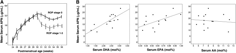
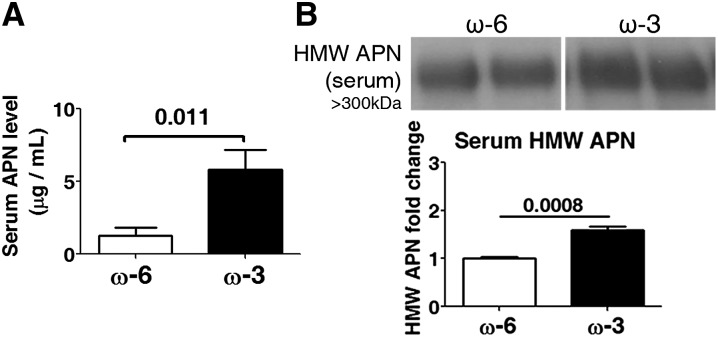
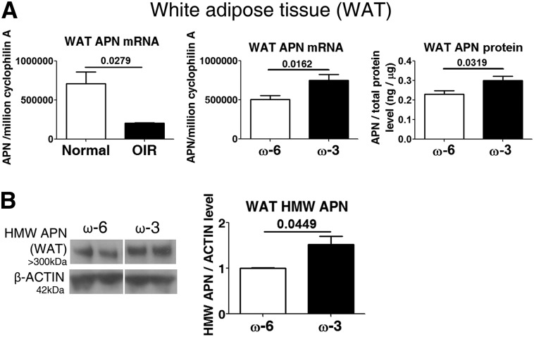
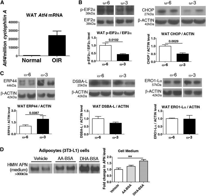
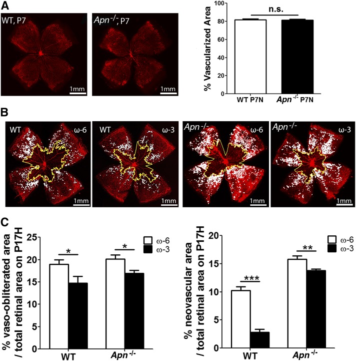
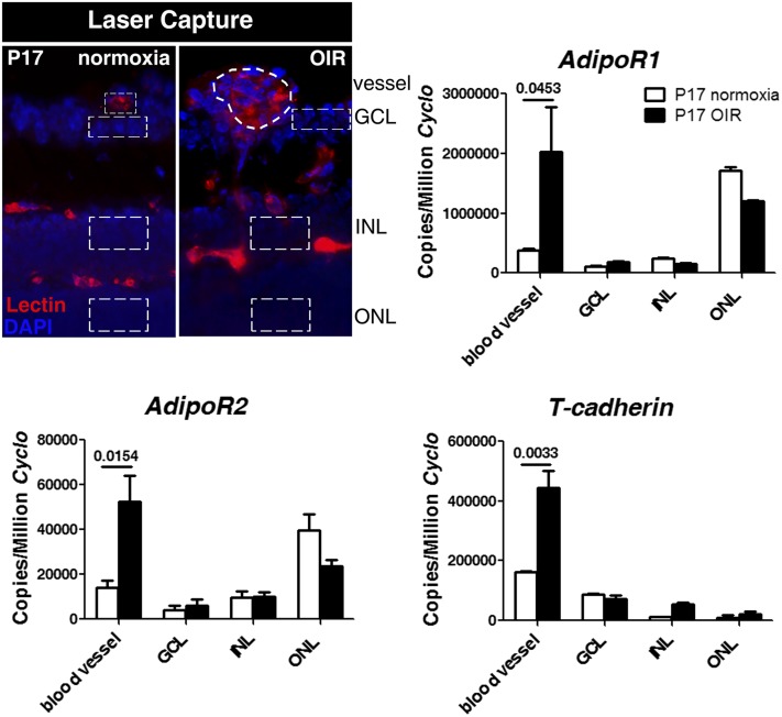
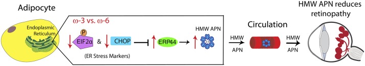
Results: We found that in preterm infants, low serum APN concentrations positively correlate with ROP, and serum APN concentrations positively correlate with serum ω-3 LCPUFA concentrations. In mouse OIR, serum total APN and bioactive high-molecular-weight APN concentrations are increased by ω-3 LCPUFA feed. White adipose tissue, where APN is produced and assembled in the endoplasmic reticulum, is the major source of serum APN. In mouse OIR, adipose endoplasmic reticulum stress is increased, and APN production is suppressed. ω-3 LCPUFA feed in mice increases APN production by reducing adipose endoplasmic reticulum stress markers. Dietary ω-3 LCPUFA suppression of neovascularization is reduced from 70% to 10% with APN deficiency. APN receptors localize in the retina, particularly to pathologic neovessels.

Conclusion: Our findings suggest that increasing APN by ω-3 LCPUFA supplementation in total parental nutrition for preterm infants may suppress ROP.

Keywords: adiponectin; endoplasmic reticulum stress; neovascularization; retinopathy of prematurity; white adipose tissue; ω-3 long-chain polyunsaturated fatty acids.


Figures

FIGURE 1
FIGURE 1
Mean (±SEM) serum DHA concentrations relate to serum APN concentrations, and lower serum APN concentrations in ROP developed in very preterm infants starting at PMA 30 wk. (A) Longitudinal serum APN concentrations in 46 very preterm infants with GA <29 wk from birth to PMA 36 wk were measured, and ROP development was monitored. n represents different independent samples. Solid line, no ROP (n = 27); dotted line, ROP (stages 1–3) (n = 19, log scale on the y axis). A t test for equality of means was used for comparison of means between 31 and 36 wk. (B) Relation of serum DHA (left, r = −0.759, P = 0.02), EPA (middle, r = −0.583, P = 0.03), and AA (right, r = −0.104, P = NS) concentrations to serum APN concentrations in very preterm infants (n = 14) with ROP development at PMA 30 wk. The Pearson correlation coefficient was used. The result was verified with Spearman’s correlation coefficient. AA, arachidonic acid; APN, adiponectin; GA, gestational age; PMA, postmenstrual age; ROP, retinopathy of prematurity.
FIGURE 2
FIGURE 2
ω-3 LCPUFAs increase total serum and HMW APN concentrations in mouse OIR. (A) ELISA for serum APN protein concentrations (n = 6). (B) Western blot for serum HMW APN (n = 4). n represents different independent samples on the same blot. Means ± SEMs are shown (unpaired t test). APN, adiponectin; HMW, high molecular weight; LCPUFA, long-chain PUFA; OIR, oxygen-induced retinopathy.
FIGURE 3
FIGURE 3
ω-3 LCPUFAs increase APN concentrations in WAT in OIR. (A) qPCR of Apn in WAT from P17 normal and OIR mice, as well as APN mRNA and protein concentrations in P17 OIR WAT (n = 6–9). (B) Western blot for HMW APN in WAT (n = 3). n represents different independent samples on the same blot. Means ± SEMs are shown (unpaired t test). APN, adiponectin; HMW, high molecular weight; LCPUFA, long-chain PUFA; mRNA, messenger RNA; OIR, oxygen-induced retinopathy; P17, postnatal day 17; qPCR, quantitative polymerase chain reaction; WAT, white adipose tissue.
FIGURE 4
FIGURE 4
ω-3 LCPUFAs modulate endoplasmic-reticulum (ER) stress in WAT. (A) qPCR of ER stress marker Atf4 in WAT from P17 normal and OIR mice (n = 3). In P17 OIR mice: protein concentrations in WAT of ER stress markers (B) p-EIF2α and CHOP (n = 4–6) and ER proteins (C) DSBA-L, ERP44, and ERO1-Lα concentrations (n = 3–7). n represents different independent samples on the same blot. Unpaired t test. (D) ω-3 LCPUFAs (DHA), not ω-6 LCPUFA (AA) modulated HMW APN in ER-stressed adipocytes. Fully differentiated 3T3-L1 cells treated with AA-BSA, DHA-BSA, or vehicle had ER stress induced with tunicamycin. HMW APN concentrations in cell medium were measured by Western blot (n = 4). n represents different independent samples on the same blot. Means ± SEMs, **P < 0.01, ANOVA. AA, arachidonic acid; APN, adiponectin; BSA, bovine serum albumin; CHOP, C/EBP homologous protein; ER, endoplasmic reticulum; HMW, high molecular weight; LCPUFA, long-chain PUFA; OIR, oxygen-induced retinopathy; P17, postnatal day 17; p-EIF2α, phospho–eukaryotic initiation factor 2α qPCR, quantitative polymerase chain reaction; WAT, white adipose tissue.
FIGURE 5
FIGURE 5
APN partly mediates the protective effects of ω-3 LCPUFAs against neovascularization. (A) Percentage of superficial vascular coverage over total retinal area at P7 in WT and Apn−/− mouse retinae (n = 10–12). Representative lectin-stained (red) retinal flat-mounts (B) and quantification (C) of vessel loss and neovascularization at P17 from WT and Apn−/− mice with either ω-3 or ω-6 LCPUFA-rich feeds from P1 to P17 (n = 7–18). n represent different independent samples. Means ± SEMs, *P < 0.05, **P < 0.01, ***P < 0.001, NS, ANOVA. Scale bar: 1 mm. APN, adiponectin; LCPUFA, long-chain PUFA; P, postnatal day; WT, wild type.
FIGURE 6
FIGURE 6
Localization of APN receptors in the LCM retinal layers and blood vessels at P17. Schematic of the LCM retinal layers (DAPI for nuclei, blue) and blood vessels (lectin, red) isolated is shown (outlined with dotted line). Expression of adipoR1, adipoR2, and T-cadherin was examined by using qPCR. n = 3. Means ± SEMs are shown (t test). APN, adiponectin; GCL, ganglion cell layer; INL, inner nuclear layer; LCM, laser-captured microdissected; OIR, oxygen-induced retinopathy; ONL, outer nuclear layer; P17, postnatal day 17; qPCR, quantitative polymerase chain reaction.
FIGURE 7
FIGURE 7
Schematic illustration of proposed pathway for ω-3 LCPUFA regulation of APN production via modulation of ER stress. In ER, activated pERK-EIF2α pathway (an unfolded protein response) in adipocytes upregulates a central transcription factor CHOP, leading to inhibition of ERP44 synthesis, an essential ER protein involved in APN assembly and secretion. Phosphorylation of EIF2α and CHOP upregulation are attenuated with ω-3 LCPUFAs compared with ω-6 LCPUFAs, thereby increasing ERP44 protein concentrations and accelerating APN assembly to a higher-order form and secretion from adipocytes into circulation. Increased serum higher-order forms of APN in turn bind their receptors adipoR1 and adipoR2 in retinal endothelial cells and macrophages, mediating protective effects of ω-3 LCPUFAs on retinal vasculature in retinopathy. APN, adiponectin; CHOP, C/EBP homologous protein; EIF2α, eukaryotic initiation factor 2α ER, endoplasmic reticulum; HMW, high molecular weight; LCPUFA, long-chain PUFA.

References

    1. Chen J, Smith LE. Retinopathy of prematurity. Angiogenesis 2007;10:133–40. - PubMed
    1. International Committee for the Classification of Retinopathy of Prematurity. The International Classification of Retinopathy of Prematurity revisited. Arch Ophthalmol 2005;123:991–9. - PubMed
    1. Palmer EA, Flynn JT, Hardy RJ, Phelps DL, Phillips CL, Schaffer DB, Tung B. Incidence and early course of retinopathy of prematurity. The Cryotherapy for Retinopathy of Prematurity Cooperative Group. Ophthalmology 1991;98:1628–40. - PubMed
    1. Stahl A, Sapieha P, Connor KM, Sangiovanni JP, Chen J, Aderman CM, Willett KL, Krah NM, Dennison RJ, Seaward MR, et al. . Short communication: PPAR gamma mediates a direct antiangiogenic effect of omega 3-PUFAs in proliferative retinopathy. Circ Res 2010;107:495–500. - PMC - PubMed
    1. Liu M, Liu F. Transcriptional and post-translational regulation of adiponectin. Biochem J 2010;425:41–52. - PubMed

Publication types

MeSH terms

LinkOut - more resources