. 2015 Apr 1;35(13):5118-27.
doi: 10.1523/JNEUROSCI.3951-14.2015.

The knockdown of αkap alters the postsynaptic apparatus of neuromuscular junctions in living mice

Affiliations

The knockdown of αkap alters the postsynaptic apparatus of neuromuscular junctions in living mice

Isabel Martinez-Pena Y Valenzuela et al. J Neurosci. .

Abstract

A muscle-specific nonkinase anchoring protein (αkap), encoded within the calcium/calmodulin kinase II (camk2) α gene, was recently found to control the stability of acetylcholine receptor (AChR) clusters on the surface of cultured myotubes. However, it remains unknown whether this protein has any effect on receptor stability and the maintenance of the structural integrity of neuromuscular synapses in vivo. By knocking down the endogenous expression of αkap in mouse sternomastoid muscles with shRNA, we found that the postsynaptic receptor density was dramatically reduced, the turnover rate of receptors at synaptic sites was significantly increased, and the insertion rates of both newly synthesized and recycled receptors into the postsynaptic membrane were depressed. Moreover, we found that αkap shRNA knockdown impaired synaptic structure as postsynaptic AChR clusters and their associated postsynaptic scaffold proteins within the neuromuscular junction were completely eliminated. These results provide new mechanistic insight into the role of αkap in regulating the stability of the postsynaptic apparatus of neuromuscular synapses.

Keywords: NMJ; imaging; knockdown; nAChR dymamics; stability; αkap.


Figures

Figure 1.
Figure 1.
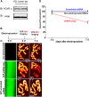
shRNA αkap significantly reduced the density of postsynaptic AChRs at the NMJ of living mice. A, Sternomastoid muscles were lysed, and receptors were labeled with BTX-biotin. Labeled AChRs were pulled down with streptavidin beads. Pull-down proteins were then probed with anti-αkap or anti-AChRα antibody. Duplicate experiments are shown. B, Experimental protocol. C, Example of NMJs from nonelectroporated or electroporated muscles with shRNA αkap or scrambled shRNA. The sternomastoid muscle of an adult mouse (WT) was electroporated with GFP-shRNA αkap or with a scrambled version of shRNA αkap. At 4 or 7 d after electroporation, AChRs were labeled with a saturating dose of BTX-Alexa Fluor 594, and superficial synapses on electroporated (green) and nonelectroporated muscle fibers from the same sternomastoid muscle were imaged. Fluorescence intensity of labeled AChRs was measured and normalized to nonelectroporated synapses. D, Graph summarizing the mean ± SD percentage of fluorescence intensity. Note that the density of AChRs was dramatically reduced in shRNA-treated muscles. ***p < 0.001.
Figure 2.
Figure 2.
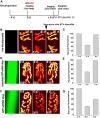
shRNA αkap impaired the insertion rate of newly synthesized AChRs. A, Labeling protocol for assessing the amount of receptor insertion. Four days after electroporation of sternomastoid muscles with shRNA-αkap or scrambled shRNA αkap, AChRs were saturated with BTX-Alexa Fluor 594, and NMJs were imaged. Six days after the initial imaging, the same NMJs were reimaged to measure the loss of fluorescence and then relabeled with a saturating dose of BTX-Alexa Fluor 594 to measure the number of new receptors that had been inserted after the initial labeling. B, Example of three views of the same image from nonelectroporated muscle. All three panels of images are displayed on the same intensity scale. D, Example of three views of the same NMJ in mice electroporated with scrambled shRNA. F, Example of three views of NMJs treated with GFP-shRNA αkap. C, E, G, Bar graphs summarizing the mean ± SE percentage of fluorescence intensity. Note that very little fluorescence was gained after BTX-Alexa Fluor 594 was added (26% of the original fluorescence), indicating that the insertion of AChR was significantly depressed in muscles electroporated with shRNA.
Figure 3.
Figure 3.
shRNA αkap decreased the contribution of recycled AChRs to the synapse. A, Scheme of the protocol used to label recycled AChRs. Seven days after electroporation, muscles were labeled with a saturating dose of BTX-biotin followed by streptavidin Alexa Fluor 594, and NMJs were imaged. Three days later, the same synapses were imaged for the second time, and then receptors were labeled with Streptavidin Alexa Fluor 594 and reimaged again. B, Examples of three views of a nonelectroporated synapse and an shRNA αkap electroporated synapse following the recycling labeling protocol described in A. C, Graph summarizing the contribution of receptor recycling to the density of an AChR. Note that the recycling of AChRs was significantly decreased in muscles treated with shRNA αkap.
Figure 4.
Figure 4.
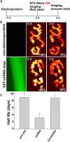
shRNA αkap enhanced the turnover rate of AChR at the postsynaptic membrane. A, Experimental protocol. The sternomastoid muscle of mice treated with shRNA αkap or scrambled shRNA were labeled with a nonsaturating dose of BTX-Alexa Fluor 594 (7 d after electroporation), and superficial synapses were imaged at time 0 and reimaged 3 d later. The total fluorescence intensity of a labeled AChR was normalized to 100% at initial imaging. B, Examples of two views of the same NMJs imaged from muscles electroporated with shRNA and nonelectroporated neighboring muscle fibers. C, Graph summarizing the half-life (days) of AChRs in NMJs treated with shRNA, nonelectroporated neighboring NMJs, or shRNA scrambled NMJs. Note that the turnover rate of AChRs is significantly higher in shRNA αkap synapses compared with nonelectroporated and scrambled synapses.
Figure 5.
Figure 5.
Disappearance of receptors from synaptic clusters in mice treated with shRNA αkap. The sternomastoid muscle was electroporated with GFP-shRNA αkap or scrambled GFP-shRNA αkap. At 7 d, muscles were labeled with BTX-Alexa Fluor 594, fixed, and imaged with a confocal microscope. A, B, Example of a confocal image of an NMJ in nonelectroporated muscle. C, D, Example of an NMJ confocal image of a muscle electroporated with scrambled shRNA αkap. E, F, Example of an NMJ confocal image of a muscle electroporated with shRNA αkap. Arrows indicate numerous perforations (holes) found in muscles treated with shRNA αkap. G, Graph showing the percentage of synapses with perforated clusters. H, Graph showing the number of perforations per NMJ in muscles electroporated with either shRNA αkap or scrambled and nonelectroporated muscles. I, J, High-resolution images of NMJs double labeled with α-BTX-Alexa Fluor 594 (AChRs, red) and VVA B4 lectin-Alexa Fluor 488 (green). K, Overlay of green and red. Note that in several synaptic clusters where receptors are missing, VVA lectin staining is still present (see white arrows), indicating that perforations form in cluster regions previously occupied by AChRs. Red arrows indicate that both AChR and VVA are missing from synaptic clusters. L, Graph showing the percentage of shRNA αkap-electroporated synapses with perforated clusters lacking α-BTX-Alexa Fluor 594 and VVA B4 lectin labeling. Note that almost 100% of NMJs are perforated after 7 d (the perforations are no longer labeled by α-BTX-Alexa Fluor 594) and that in 50% of those synapses VVA B4 lectin is still present. Scale bars, 5 μm.
Figure 6.
Figure 6.
Neuromuscular junction disassembly in muscles treated with shRNA αkap. The sternomastoid muscle was electroporated with shRNA αkap and rapsyn-GFP, α-syntrophin-GFP, α-dystrobrevin GFP, or GFP-CaMKIIβM; and 7 d after electroporation, muscles were labeled with BTX-Alexa Fluor 594 and fixed, and then synapses were imaged. A–C, Example of a high-resolution confocal image of a NMJ expressing rapsyn-GFP and labeled with BTX-Alexa Fluor 594. Note that rapsyn-GFP colocalized perfectly with receptors at synaptic sites, except in regions where receptors have been lost (see arrows), indicating that rapsyn and AChRs are removed simultaneously. D–L, Examples of a high-resolution confocal image of a NMJ expressing α-dystrobrevin-GFP (αDB-GFP; D–F), α-syntrophin-GFP (G–I), and GFP-CaMKIIβM (J–L). Note that α-syntrophin, α-dystrobrevin, and CaMKIIβM are no longer present at some perforated synaptic clusters (red arrows), but not in others (white arrows), suggesting that AChRs and these molecules have different rates of removal. M–R, Seven days (M–O) and 21 d (P–R) after electroporation with shRNA αkap, muscles were fixed and stained with antibodies against Sv2/neurofilament (green) and BTX-Alexa Fluor 594. Note that synaptic nerve terminal branches remain visible at synaptic sites where receptors are lost at 7 d (white arrows), but they are removed by 21 d (red arrows); asterisks show areas of synaptic clusters where receptors are faint or completely missing. Scale bars, 5 μm.

Similar articles

Cited by

References

    1. Adams ME, Kramarcy N, Krall SP, Rossi SG, Rotundo RL, Sealock R, Froehner SC. Absence of alpha-syntrophin leads to structurally aberrant neuromuscular synapses deficient in utrophin. J Cell Biol. 2000;150:1385–1398. doi: 10.1083/jcb.150.6.1385. - DOI - PMC - PubMed
    1. Akaaboune M, Culican SM, Turney SG, Lichtman JW. Rapid and reversible effects of activity on acetylcholine receptor density at the neuromuscular junction in vivo. Science. 1999;286:503–507. doi: 10.1126/science.286.5439.503. - DOI - PubMed
    1. Bayer KU, Löhler J, Harbers K. An alternative, nonkinase product of the brain-specifically expressed Ca2+/calmodulin-dependent kinase II alpha isoform gene in skeletal muscle. Mol Cell Biol. 1996;16:29–36. - PMC - PubMed
    1. Bayer KU, Harbers K, Schulman H. alphaKAP is an anchoring protein for a novel CaM kinase II isoform in skeletal muscle. EMBO J. 1998;17:5598–5605. doi: 10.1093/emboj/17.19.5598. - DOI - PMC - PubMed
    1. Bayer KU, De Koninck P, Schulman H. Alternative splicing modulates the frequency-dependent response of CaMKII to Ca(2+) oscillations. EMBO J. 2002;21:3590–3597. doi: 10.1093/emboj/cdf360. - DOI - PMC - PubMed

Publication types

MeSH terms

Substances

LinkOut - more resources