. 2015 Apr 1;35(13):5221-32.
doi: 10.1523/JNEUROSCI.4650-14.2015.

Definition of a molecular pathway mediating α-synuclein neurotoxicity

Affiliations

Definition of a molecular pathway mediating α-synuclein neurotoxicity

Jacqueline Burré et al. J Neurosci. .

Erratum in

Abstract

α-Synuclein physiologically chaperones SNARE-complex assembly at the synapse but pathologically misfolds into neurotoxic aggregates that are characteristic for neurodegenerative disorders, such as Parkinson's disease, and that may spread from one neuron to the next throughout the brain during Parkinson's disease pathogenesis. In normal nerve terminals, α-synuclein is present in an equilibrium between a cytosolic form that is natively unfolded and monomeric and a membrane-bound form that is composed of an α-helical multimeric species that chaperones SNARE-complex assembly. Although the neurotoxicity of α-synuclein is well established, the relationship between the native conformations of α-synuclein and its pathological aggregation remain incompletely understood; most importantly, it is unclear whether α-synuclein aggregation originates from its monomeric cytosolic or oligomeric membrane-bound form. Here, we address this question by introducing into α-synuclein point mutations that block membrane binding and by then assessing the effect of blocking membrane binding on α-synuclein aggregation and neurotoxicity. We show that membrane binding inhibits α-synuclein aggregation; conversely, blocking membrane binding enhances α-synuclein aggregation. Stereotactic viral expression of wild-type and mutant α-synuclein in the substantia nigra of mice demonstrated that blocking α-synuclein membrane binding significantly enhanced its neurotoxicity in vivo. Our data delineate a folding pathway for α-synuclein that ranges from a physiological multimeric, α-helical, and membrane-bound species that acts as a SNARE-complex chaperone over a monomeric, natively unfolded form to an amyloid-like aggregate that is neurotoxic in vivo.

Keywords: Parkinson's disease; aggregation; alpha-synuclein; membrane binding.


Figures

Figure 1.
Figure 1.
Design of lipid-binding deficient mutants of α-synuclein. A, α-Synuclein domain structure (red, lipid-binding domain; blue, protein-interaction domain), with lipid-binding deficient mutations marked in red and 11-mer sequences highlighted with black boxes. B, SDS-PAGE analysis of purified recombinant mutant α-synuclein (5 μg/lane), stained with Coomassie Brilliant Blue. C, D, Lipid binding of wild-type and mutant α-synuclein. C, Recombinant α-synuclein was incubated with negatively charged liposomes (composition: 30% phosphatidylserine (PS) and 70% phosphatidylcholine (PC)) and subjected to a flotation assay. Eight fractions were collected from top to bottom of the flotation gradient, and equal volumes of each fraction were separated by SDS-PAGE and immunoblotted for α-synuclein. D, The top two fractions were defined as lipid bound and quantitated as percentage of total α-synuclein (means ± SEMs; **p < 0.001 by Mann–Whitney U test; n = 3). E, Analysis of the effect of α-synuclein mutations on membrane-binding induced α-synuclein multimerization. Recombinant α-synuclein was incubated with negatively charged liposomes (composition: 30% PS, 70% PC) and exposed to increasing concentrations of the chemical crosslinker glutaraldehyde (concentration: 0–0.5%). Equal volumes of crosslinked proteins were analyzed by immunoblotting. Arrowheads indicate α-synuclein multimers.
Figure 2.
Figure 2.
Aggregation of α-synuclein in vitro. A, B, Recombinant wild-type α-synuclein or mutant α-synuclein unable to bind to liposomes were incubated in buffer (left panels) or in the presence of charged liposomes (composition: 30% PS, 70% PC; right panels) at 37°C and 300 rpm. At the indicated time points, aggregation of α-synuclein was assessed by light microscopy.
Figure 3.
Figure 3.
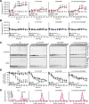
In vitro aggregation of α-synuclein (αSyn). A, B, Amyloid formation of α-synuclein. Recombinant α-synuclein was incubated as described for Figure 2. At the indicated time points, wild-type and mutant α-synuclein were analyzed for amyloid fibril formation in buffer (A) or in the presence of charged liposomes (B), using the dye K114 (means ± SEMs; *p < 0.05, **p < 0.01, ***p < 0.001 by Mann–Whitney U test; ##p < 0.01, ###p < 0.001 by two-way ANOVA; n = 7; n.s., not significant). C, D, Analysis of loss of monomer of α-synuclein. At the indicated time points, the same volumes of wild-type and mutant α-synuclein aggregating in buffer or in the presence of charged liposomes were analyzed by immunoblotting, and loss of monomer was quantitated (means ± SEMs; **p < 0.01 by Mann–Whitney U test; ###p < 0.001 by two-way ANOVA; n = 7; n.s., not significant). E, Analysis of wild-type and mutant α-synuclein by gel filtration. At the indicated time points, 90 μl of aggregating recombinant wild-type and mutant α-synuclein were analyzed by gel filtration. Visible aggregates were removed before loading via centrifugation to avoid clogging of the gel filtration column.
Figure 4.
Figure 4.
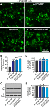
Aggregation and toxicity of α-synuclein in HEK293T cells. A, B, Aggregation of wild-type and mutant α-synuclein in HEK293T cells transfected with equal amounts of expression vectors encoding wild-type and mutant α-synuclein. A, Two days after transfection, cells were fixed and immunostained with antibodies against the myc-epitope. B, The number of immunopositive aggregates per field was quantitated and compared with wild-type levels (means ± SEMs; ***p < 0.001 by Mann–Whitney U test; n = 3 independent cultures). C, D, Expression of wild-type and mutant α-synuclein (α-Syn) in HEK293T cells. Two days after transfection, expression levels were analyzed by immunoblotting with antibodies against the myc-epitope (C), normalized to β-actin levels, and quantitated as percentage of wild-type levels (D; means ± SEMs; n = 4 independent cultures). E, Metabolic activity of HEK293T cells transfected with wild-type and mutant α-synuclein. Two days after transfection, cells were subjected to an MTT assay. Data were normalized to wild-type α-synuclein levels (means ± SEMs; *p < 0.05 by Mann–Whitney U test; n = 6 independent cultures).
Figure 5.
Figure 5.
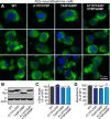
Aggregation and toxicity of α-synuclein in N2a neuroblastoma cells. A, Expression of wild-type and mutant α-synuclein in N2a neuroblastoma cells. Two days after transfection, cells were fixed and immunostained with antibodies against the myc epitope. Nuclei were visualized using DAPI. B, C, Expression of wild-type and mutant α-synuclein (α-Syn) in N2a neuroblastoma cells. Two days after transfection, expression levels were analyzed by immunoblotting with antibodies against the myc epitope (B), normalized to β-actin levels, and quantitated as percentage of wild-type levels (C; means ± SEMs; n = 6 independent cultures). D, Metabolic activity of N2a neuroblastoma cells transfected with wild-type and mutant α-synuclein. Two days after transfection, cells were subjected to an MTT assay. Data were normalized to wild-type α-synuclein levels (means ± SEMs; *p < 0.05, **p < 0.01 by Mann–Whitney U test; n = 6 independent cultures).
Figure 6.
Figure 6.
Lentiviral expression of wild-type and mutant α-synuclein in dopaminergic substantia nigra neurons. A, Schematic overview of the stereotactic injection experiments. Wild-type mice (40–45 d old) were stereotactically and unilaterally injected into the substantia nigra (left). Mice were monitored every 5 d from 10 to 45 d after injection, when mice were killed for histochemical analysis (right). B, C, Beam-walk assay. B, Motor defects were assayed using the beam-walk task in which foot slips on a beam walk were measured. Three rounds of beam walk were analyzed per session. Averaged foot slips were recorded. C, Quantitation of average foot slips at 45 d after injections. Data are means ± SEMs (*,#p < 0.05 by Mann–Whitney U test; n = 5 mice).
Figure 7.
Figure 7.
Motor impairments of mice injected with lentiviral particles expressing α-synuclein variants. A, Representative traces of force-plate analyses. Mice were injected and analyzed as described for Figure 5. B–I, Analysis of spatial confinement, total and continuous distance traveled, and low mobility bouts, as calculated from the force-plate data obtained with multiple identically injected mice. Every 5 d, mice were subjected to behavioral analyses (B, D, F, H), and data were plotted as the difference at 45 d after injection (C, E, G, I). Data are means ± SEMs (*,#p < 0.05, **,##p < 0.01 by Mann–Whitney U test; n = 5 mice).
Figure 8.
Figure 8.
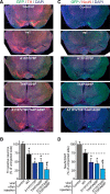
Neuron loss in mice expressing α-synuclein (αSyn) mutants in the substantia nigra. A, C, Control lentiviruses (control) or lentiviruses expressing wild-type or mutant α-synucleins were stereotactically and unilaterally injected into the substantia nigra of 40- to 45-d-old mice. Forty-five days after injection, injected areas were immunostained for either tyrosine hydroxylase (TH; left) or NeuN (right) and DAPI (blue). IRES-driven GFP marks the injection site. B, D, The density of dopaminergic neurons was quantitated by immunostaining for TH (B), and the density of NeuN-positive (non-dopaminergic) neurons was quantitated by immunostaining for NeuN (D). Data are means ± SEMs (*,#p < 0.05 by Mann–Whitney U test; n = 5 mice).
Figure 9.
Figure 9.
Schematic of α-synuclein conformations associated with its physiological function and pathological activities. Soluble α-synuclein is natively unstructured and monomeric. After binding to highly curved membranes such as synaptic vesicles, α-synuclein undergoes a conformational change and folds into a broken amphiphathic α-helix, which is associated with multimerization and mediates its SNARE-complex chaperoning function. Under pathological conditions, soluble α-synuclein forms β-sheet-like oligomers (protofibrils), which convert into amyloid fibrils and eventually deposit into Lewy bodies. Protofibrils and fibrils may propagate from neuron to neuron in PD and Lewy body dementia and from glia to glia in multiple system atrophy.

References

    1. Arawaka S, Saito Y, Murayama S, Mori H. Lewy body in neurodegeneration with brain iron accumulation type 1 is immunoreactive for alpha-synuclein. Neurology. 1998;51:887–889. doi: 10.1212/WNL.51.3.887. - DOI - PubMed
    1. Azeredo da Silveira S, Schneider BL, Cifuentes-Diaz C, Sage D, Abbas-Terki T, Iwatsubo T, Unser M, Aebischer P. Phosphorylation does not prompt, nor prevent, the formation of alpha-synuclein toxic species in a rat model of Parkinson's disease. Hum Mol Genet. 2009;18:872–887. doi: 10.1093/hmg/ddn417. - DOI - PubMed
    1. Barenholz Y, Gibbes D, Litman BJ, Goll J, Thompson TE, Carlson RD. A simple method for the preparation of homogeneous phospholipid vesicles. Biochemistry. 1977;16:2806–2810. doi: 10.1021/bi00631a035. - DOI - PubMed
    1. Breydo L, Wu JW, Uversky VN. Alpha-synuclein misfolding and Parkinson's disease. Biochim Biophys Acta. 2012;1822:261–285. doi: 10.1016/j.bbadis.2011.10.002. - DOI - PubMed
    1. Burré J, Sharma M, Tsetsenis T, Buchman V, Etherton MR, Südhof TC. Alpha-synuclein promotes SNARE-complex assembly in vivo and in vitro. Science. 2010;329:1663–1667. doi: 10.1126/science.1195227. - DOI - PMC - PubMed

Publication types

LinkOut - more resources