. 2015 May 10;6(13):10880-92.
doi: 10.18632/oncotarget.3534.

PIM1 regulates glycolysis and promotes tumor progression in hepatocellular carcinoma

Affiliations

PIM1 regulates glycolysis and promotes tumor progression in hepatocellular carcinoma

Carmen Oi-ning Leung et al. Oncotarget. .

Abstract

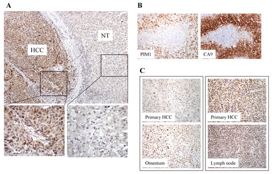
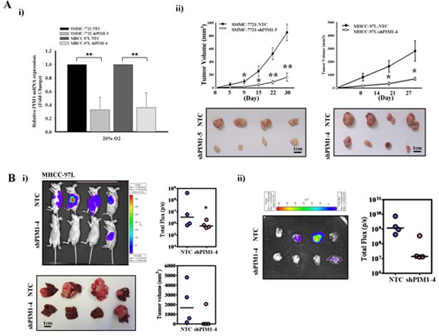
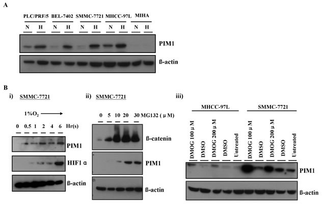
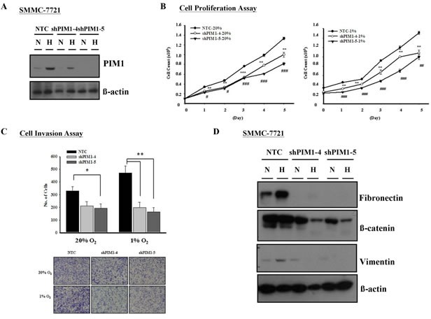
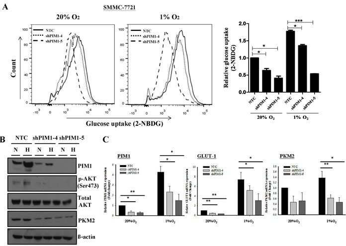
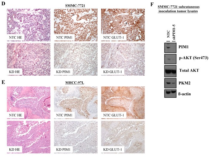
Hepatocellular carcinoma (HCC) is characteristically one of the most rapidly proliferating tumors which outgrows functional blood supply and results in regional oxygen deprivation. Overexpression of PIM1, a serine/threonine kinase, has been identified recently in human cancers. Knowledge on PIM1 in HCC is however, scarce. By immunohistochemical analysis on 56 human primary HCC samples, we observed overexpression of PIM1 in 39% of the cases. In two independent cohorts of paired primary and extra-hepatic metastatic HCC tissues, PIM1 expression was higher (p=0.002) in the extra-hepatic metastatic HCC tissues as compared with the corresponding primary HCCs. PIM1 was markedly up-regulated in multiple HCC cell lines in hypoxic condition (1% O2) versus normoxia (20% O2). Silencing of PIM1 suppressed HCC cell invasion in vitro as compared to non-target control, and decreased HCC cell proliferation in vitro and tumor growth and metastatic potential in vivo. Knockdown of PIM1 significantly reduced glucose uptake by HCC cells and was associated with decreased levels of p-AKT and key molecules in the glycolytic pathway. Taken together, PIM1 is up-regulated by hypoxia in HCC and promotes tumor growth and metastasis through facilitating cancer cell glycolysis. Targeting PIM1 may have potential role in the management of HCC.

Keywords: HCC; PIM1; metastasis.


Figures

Figure 1
Figure 1
(A) Expression of PIM1 in HCC tissues by immunohistochemistry. PIM1 was overexpressed in HCC. Positive nuclear and cytoplasmic staining was observed in the HCC tumors. No staining was present in the corresponding non-tumorous (NT) tissues. (B) Immunohistochemical staining of PIM1 and CA9 in HCC tissues. (C) PIM1 immunohistochemical expression in paired primary and extra-hepatic metastatic HCC tissues. Left panel - PIM1 staining was negative in the primary HCC tissue but was positive in the corresponding metastatic HCC tissue in the omentum; Right panel - PIM1 staining was positive in both primary HCC and the corresponding metastatic HCC tissue in the lymph node.
Figure 2
Figure 2. Silencing of PIM1 suppressed the tumor volume and metastatic ability in vivo
(A) i) Quantitative PCR analysis of PIM1 expression in NTC and PIM1 stable knockdown clones in SMMC-7721 (shPIM1-5) and MHCC-97L (shPIM1-4), respectively. ii) Subcutaneously inoculation of NTC and shPIM1-5 (SMMC-7721) or shPIM1-4 (MHCC-97L). Tumor growth volume upon inoculation measured with calipers; tumors were excised from the two groups of mice. (B) i) Xenogen imaging of nude mice orthotopically implanted with tumors derived from MHCC-97L-NTC and shPIM1-4 at 6th week upon implantation, with tumor incidence rate indicated. Livers from the two groups of mice at 6th week upon implantation, with tumor volume indicated respectively. ii) Ex-vivo imaging of lung from the two groups of mice, with pulmonary metastasis rate indicated.
Figure 3
Figure 3. PIM1 expression was up-regulated by hypoxia, which was HIF-1α independent
(A) PIM1 expression in HCC cell lines and MIHA. N: normoxia (20% O2) H: hypoxia (1% O2). (B) i) PIM1 protein expression was up-regulated at around 30 minutes and preceded that of HIF-1α in SMMC-7721. ii) Treatment of SMMC-7721 with proteasome inhibitor MG132 in normoxia stabilized PIM1 protein. iii) Treatment with propyl hydroxylase inhibitor DMOG in SMMC-7721 and MHCC-97L also stabilized PIM1 protein.
Figure 4
Figure 4. Silencing of PIM1 inhibited cell proliferation and invasiveness in HCC (SMMC-7721)
(A) Western blot analysis of PIM1 expression in PIM1 stable knockdown and NTC clones. (B) Silencing of PIM1 suppressed cell proliferation in both normoxic and hypoxia conditions (* NTC vs shPIM1-4; # NTC vs shPIM1-5; */# p<0.05; **/## p<0.01; ***/### p<0.001). (C) Upper panel - Knockdown of PIM1 inhibited cell invasiveness in normoxic and hypoxic conditions (20% O2: NTC vs shPIM1-4 p=0.053; NTC vs shPIM1-5 p=0.030; 1% O2: NTC vs shPIM1-4 p=0.008; NTC vs shPIM1-5 p=0.003). Lower panel - Representative pictures of transwell after staining with 1% crystal violet. (D) Expression of epithelial-mesenchymal markers in PIM1 stable knockdown clones shPIM1-4 and shPIM1-5 of SMMC-7721.
Figure 5
Figure 5. Knockdown of PIM1 suppressed the glycolytic metabolism in SMMC-7721, possibly through the regulation of p-AKT
(A) Glucose uptake experiment using 2-NBDG flow cytometry analyses (left panel) and relative glucose uptake (right panel). (B) Expression of p-AKT, total AKT and pyruvate kinase M2 (PKM2) in SMMC-7721 stable PIM1 knockdown clones shPIM1-4 and shPIM1-5. N: normoxia (20% O2) H: hypoxia (1% O2). (C) Expression of GLUT-1 and PKM2 by qPCR in PIM1 stable knockdown clones of SMMC-7721. (D) Immunohistochemical staining of PIM1 and GLUT-1 of HCC tissues in NTC and PIM1 knockdown (KD) clones from subcutaneous inoculation (SMMC-7721) and (E) orthotopic implantation (MHCC-97L) of nude mice. (F) Expression of PIM1, p-AKT, total AKT and PKM2 of SMMC-7721 tumor lysates from NTC and shPIM1-5 after subcutaneous inoculation in nude mice.

Similar articles

Cited by

References

    1. Semenza GL. Hypoxia-inducible factors in physiology and medicine. Cell. 2012;148:399–408. - PMC - PubMed
    1. Epstein AC, Gleadle JM, McNeill LA, Hewitson KS, O'Rourke J, Mole DR, Mukherji M, Metzen E, Wilson MI, Dhanda A, Tian YM, Masson N, Hamilton DL, et al. C. elegans EGL-9 and mammalian homologs define a family of dioxygenases that regulate HIF by prolyl hydroxylation. Cell. 2001;107:43–54. - PubMed
    1. Kaelin WG, Jr, Ratcliffe PJ. Oxygen sensing by metazoans: the central role of the HIF hydroxylase pathway. Mol Cell. 2008;30:393–402. - PubMed
    1. Lu X, Kang Y. Hypoxia and hypoxia-inducible factors: master regulators of metastasis. Clin Cancer Res. 2010;16:5928–35. - PMC - PubMed
    1. Bangoura G, Liu ZS, Qian Q, Jiang CQ, Yang GF, Jing S. Prognostic significance of HIF-2alpha/EPAS1 expression in hepatocellular carcinoma. World J Gastroenterol. 2007;13:3176–82. - PMC - PubMed

Publication types

MeSH terms

Substances