. 2015 Apr 1;7(281):281ra45.
doi: 10.1126/scitranslmed.aaa5171.

Neuregulin stimulation of cardiomyocyte regeneration in mice and human myocardium reveals a therapeutic window

Affiliations

Neuregulin stimulation of cardiomyocyte regeneration in mice and human myocardium reveals a therapeutic window

Brian D Polizzotti et al. Sci Transl Med. .

Abstract

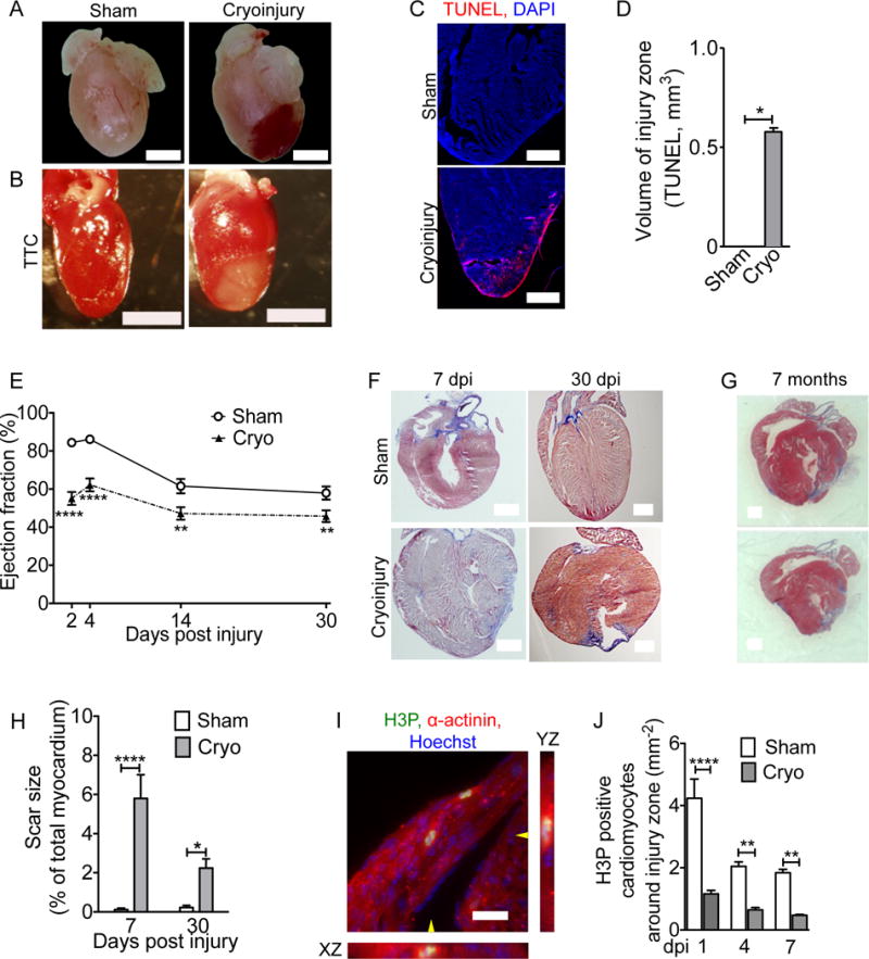
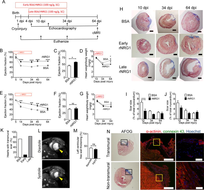
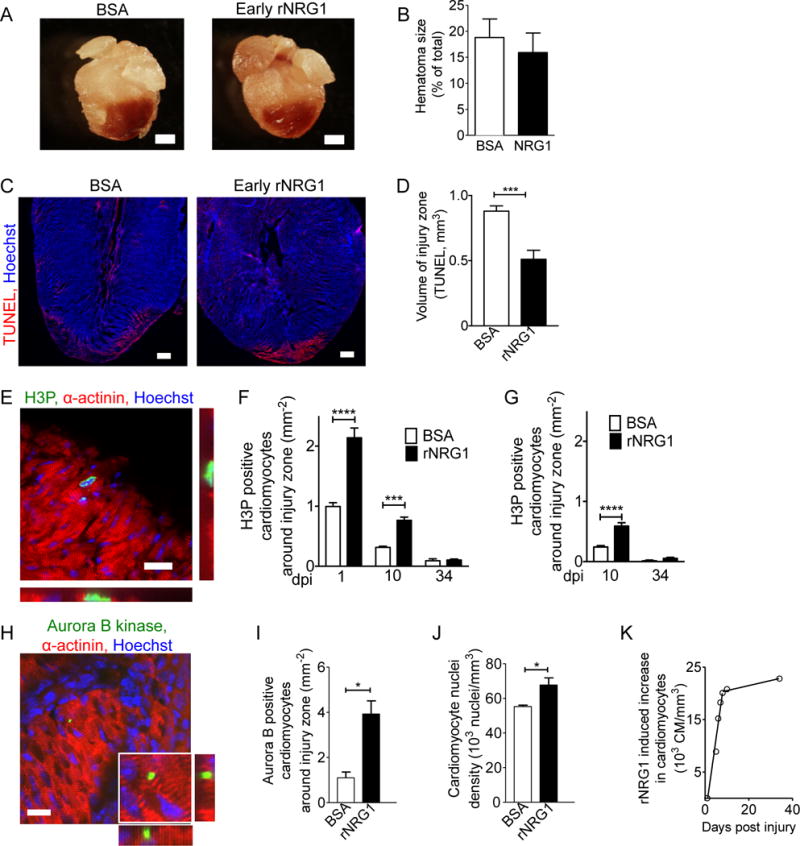
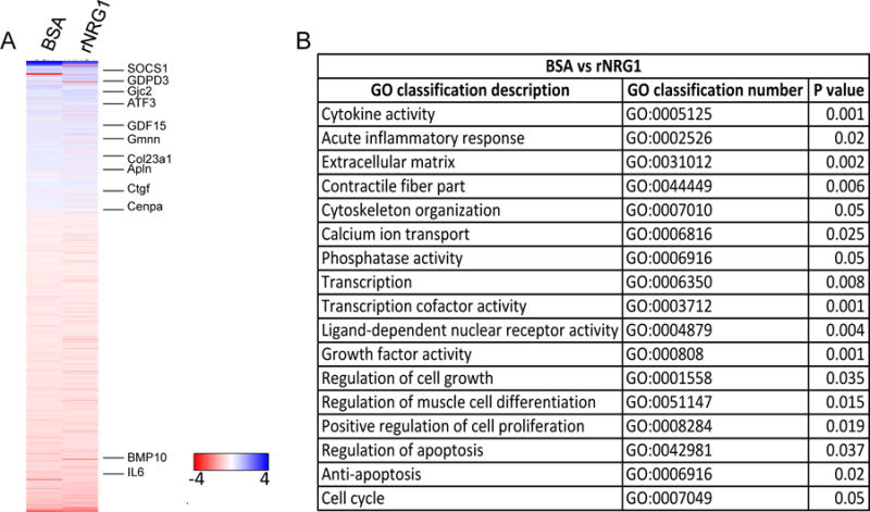
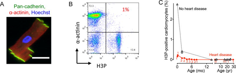
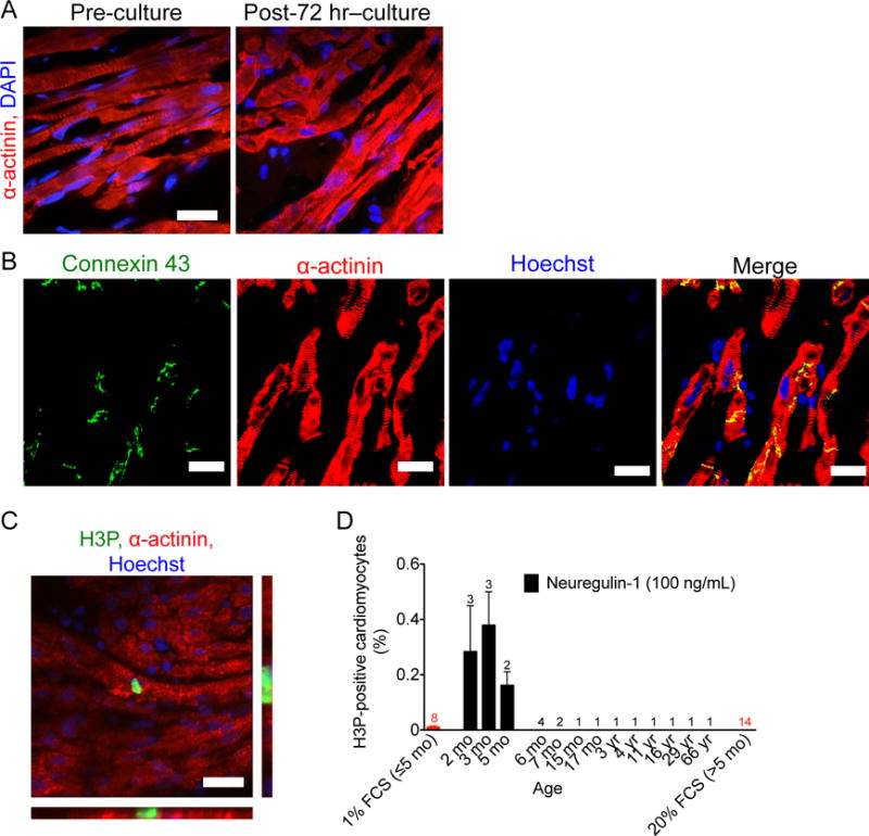
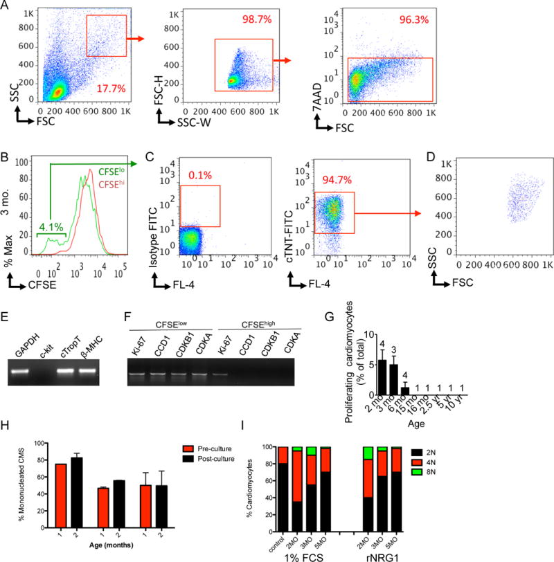
Therapies developed for adult patients with heart failure have been shown to be ineffective in pediatric clinical trials, leading to the recognition that new pediatric-specific therapies for heart failure must be developed. Administration of the recombinant growth factor neuregulin-1 (rNRG1) stimulates regeneration of heart muscle cells (cardiomyocytes) in adult mice. Because proliferation-competent cardiomyocytes are more abundant in growing mammals, we hypothesized that administration of rNRG1 during the neonatal period might be more effective than in adulthood. If so, neonatal rNRG1 delivery could be a new therapeutic strategy for treating heart failure in pediatric patients. To evaluate the effectiveness of rNRG1 administration in cardiac regeneration, newborn mice were subjected to cryoinjury, which induced myocardial dysfunction and scar formation and decreased cardiomyocyte cell cycle activity. Early administration of rNRG1 to mice from birth to 34 days of age improved myocardial function and reduced the prevalence of transmural scars. In contrast, administration of rNRG1 from 4 to 34 days of age only transiently improved myocardial function. The mechanisms of early administration involved cardiomyocyte protection (38%) and proliferation (62%). We also assessed the ability of rNRG1 to stimulate cardiomyocyte proliferation in intact cultured myocardium from pediatric patients. rNRG1 induced cardiomyocyte proliferation in myocardium from infants with heart disease who were less than 6 months of age. Our results identify an effective time period within which to execute rNRG1 clinical trials in pediatric patients for the stimulation of cardiomyocyte regeneration.


Conflict of interest statement

Competing interests: The authors declare no conflicting financial interests.

Figures

Figure 1
Figure 1. Cryoinjury induces cell death, myocardial dysfunction, and decreased cardiomyocyte cell cycle activity in neonatal mice
Mice underwent sham surgery or cryoinjury on day of life 1 (P1). (A) Hematoma at the injury site. (B) Vital staining with triphenyltetrazolium chloride (TTC) shows injury zone. (C,D) Myocardial cell death visualized by TUNEL staining. (E) Cryoinjury induces a sustained decrease in the ejection fraction. (F,G) AFOG-stained sections show that scar (blue) is formed within 7 dpi and present 30 days later (F). (G) Cyoinjury-induced scars, visualized on two sections of the same heart (500 μm apart) by Masson Trichrome staining, persist to 7 months after injury. (H) Quantification of scar size. (I) Two cardiomyocytes in M-phase visualized with antibodies against phosphorylated histone H3 (H3P) and α-actinin. The position of orthogonal reconstructions of the cardiomyocyte in the center are indicated with yellow arrowheads. (J) Quantification of M-phase cardiomyocytes in the region around the injury zone shows significant and sustained reduction after cryoinjury. Scale bars 1 mm (A,B,F,G), 20 μm (C,I). Statistical test by t-test (D) and ANOVA Bonferroni’s Multiple Comparison Test (E, H, J) * P < 0.05, ** P < 0.01, *** P < 0.001, **** P < 0.0001.
Figure 2
Figure 2. Early administration of rNRG1 improves myocardial function and structure
(A) Experimental design of mouse pre-clinical trials. (B–K) Mice underwent cryoinjury on day 1 of life (P1) and were treated with BSA or rNRG1 from day of birth (P0, early administration, B–D), or from P5 (late administration, E–G). (B,C) Prolonged improvement in myocardial function after early administration shown by echocardiography (B) and cMRI at 64 dpi (C). (E,F) Late administration of rNRG1 resulted in transient improvement of myocardial function measured by echocardiography (E) and cMRI at 64 dpi (F). (D,G) Indexed heart weights showed early rNRG1 administration reduced cardiac hypertrophy at 64 dpi. (H) Time-series of AFOG-stained section shows scar (blue) is formed within 10 dpi and still present at 64 dpi. Note transmural scars after cryoinjury in BSA and late rNRG1 treatment groups. (I,J) Quantification of scar size after AFOG-staining shows transient and significant scar reduction after early rNRG1 administration (I). (K) Early administration reduces the percent of transmural scars at 34 and 64 dpi. (L) Non-transmural injury site thickens in systole (64 dpi, early administration) (M) Relative thickening of non-transmural scars is similar to remote LV free wall myocardium. (N) Transmural and non-transmural scars were identified by AFOG sections (left panels). Black rectangles indicate photomicrographs shown in the middle panels. Non-transmural scars have cardiomyocytes connected by gap junctions visualized with Connexin 43 staining (64 dpi, early administration, middle and right panels). Yellow squares indicate zoomed areas (Right panels). Scale bars 1 mm (H), 500 μm (N, center panel), 50 μm (N, far right). dpi: days post injury; BSA: bovine serum albumin; SC: subcutaneous injection: P0: day of birth. Statistical significance was tested with t-test (C,F,M) and ANOVA Bonferroni's Multiple Comparison Test (B,D,E,G,I,J,K) * P < 0.05, ** P < 0.01, *** P < 0.001, **** P < 0.0001.
Figure 3
Figure 3. Early administration of rNRG1 reduces myocardial death and stimulates cardiomyocyte proliferation
(A,B) Hematomas are present at the zone of injury at 1 dpi (A). Hematoma size quantification shows no change after early administration of rNRG1 (B). (C,D) Photomicrographs (C) and quantification (D) of myocardial cell death visualized by TUNEL staining at 1 dpi after early administration. (E) Cardiomyocytes in M-phase were visualized with an antibody against phosphorylated histone H3 (H3P). (F,G) H3P-positive cardiomyocytes were quantified around injury zone after early (F) and late (G) administration. Treatment with rNRG1 increases cardiomyocyte cell cycle activity, and early administration captures the regenerative phase (F). (H,I) Cardiomyoctyes in cytokinesis were visualized with an antibody against Aurora B kinase (H) and quantified around the injury zone after early administration at 1 dpi (I). (J) Cardiomyocyte nuclear density is increased after early administration of rNRG1 (34 dpi). (K) Early administration of rNRG1 increases the cardiomyocyte density by ~62,000 cardiomyocytes/mm3 within the first 8 days, compared to BSA controls. Scale bars 1 mm (A), 20 μm (C,E,H). Statistical significance was tested with t-test (B,D,I,J) and ANOVA (F,G) * P < 0.05, *** P < 0.001, **** P < 0.0001.
Figure 4
Figure 4. rNRG1 acts via ErbB4 on cardiomyocytes in neonatal mouse hearts in vivo
(A–C) Experiments were performed in α-MHC-Mer-Cre-Mer+/+; ErbB4F/wt (control) and in α-MHC-Mer-Cre-Mer+/+; ErbB4F/F (test). rNRG1 or BSA was administered from day of birth (P0) until P12. ErbB4 inactivation was induced with tamoxifen administration on days P1–3, which caused a significant down-regulation of ErbB4 mRNA levels (A). Representative example of a cardiomyocyte in M-phase with orthogonal reconstructions (H3P, B). rNRG1 increased cardiomyocyte cell cycle activity in ErbB4F/wt, but not in ErbB4F/F mice (C). Statistical test by t-test (A) and ANOVA Bonferroni's Multiple Comparison Test (C) * P < 0.05, scale bar 50 μm.
Figure 5
Figure 5. Cryoinjury and rNRG1 administration induce gene regulation patterns that parallel structural and functional changes
Mice underwent cryoinjury on day 1 of life (P1) and were treated with BSA or rNRG1 according to the early administration protocol. Expression profiling was performed at 10 dpi with 5 mice per group and normalized to sham (n = 5). (A) Heat map shows 622 genes that were significantly regulated between BSA and rNRG1 treated mice (P<0.05). Selected genes discussed in the text are indicated. The color chart indicates fold change of expression using a log2 scale. (B) Functional annotation clustering of differentially expressed genes shows significant regulation of multiple pathways by rNRG1. GO, gene ontology.
Figure 6
Figure 6. Pediatric patients with heart disease show decreased cardiomyocyte cell cycle activity
Cardiomyocytes from patients were isolated, stained, and analyzed by flow cytometry. (A) Isolated human cardiomyoctes were intact as evidenced by staining with antibodies against pan-cadherin and α-actinin. (B) Representative double marker plot of a 3 month old patient showing flow cytometry analysis of cardiomyocyte cell cycle activity using cardiomyocyte (α-actinin) and cell cycle markers (H3P). (C) Summary graph shows that patients with heart disease exhibit decreased cycling compared to age-matched controls without heart disease. Numbers of patients per data point are indicated in red, no heart disease were 1 patient per data point. Circles represent RV and triangles LV samples. Scale bar: 50 μm.
Figure 7
Figure 7. rNRG1 stimulates cardiomyocyte cycling in myocardium from infants with heart disease younger than 6 months of age
For organotypic culture, chunks of myocardium were maintained in the presence of 1% FCS or rNRG1 for 3 days, fixed, and analyzed by immunofluorescence microscopy. (A,B) Organotypic culture for 3 days does not change microscopic architecture (A). Gap junctions, electromechanical connections, were identified by connexin 43 staining were present after 72 hours of organotypic culture (B). (C,D) rNRG1 stimulates cardiomyocytes to enter M-phase in a 2-month old patient with ToF (C). Quantitative analysis showed that rNRG1 increased M-phase cardiomyocytes in an age-dependent manner (D). Numbers of patients per data point are indicated (D). Scale bars 20 μm (A,C), 50 μm (B).
Figure 8
Figure 8. rNRG1 stimulates cardiomyocyte proliferation in myocardium from patients with heart disease younger than 6 months of age
Organotypic cultures of human myocardium were metabolically labeled with CFSE and then maintained in the presence of 1% FBS or rNRG1 for 3 days. Cardiomyocytes were analyzed and isolated by FACS. (A) FACS strategy for enrichment by size (left panel), doublet discrimination (middle panel), and viability (right panel). (B) Flow cytometry analysis of a 3 months-old reveals a CFSElo population of 4.1%. (CD) After fixation, this population was stained with isotype control (C, left panel) and antibodies against troponin T (C, right panel). Analysis by flow cytometry shows that 94.7% of this population were cardiomyocytes (C, right panel) with a forward and side scatter characteristics (D) similar to (A). (E,F) RT-PCR showed that CFSElo cardiomyocytes expressed markers of mature differentiated cardiomyocytes (E) and cell-cycle associated genes (F). (G) Graph of proportion of CFSElo populations shows that stimulation of cardiomyocyte proliferation in patients with heart disease is age-dependent. Numbers of patients per data point are indicated. (H,I) Laser scanning cytometry shows that administration of rNRG1 in organotypic culture did not change the overall percentage of mononucleated cardiomyocytes (H) or the ploidy pattern of mononucleated cardiomyocytes (I).

References

    1. Tennant PW, Pearce MS, Bythell M, Rankin J. 20-year survival of children born with congenital anomalies: a population-based study. Lancet. 2010;375:649. - PubMed
    1. Hoffman JI, Kaplan S. The incidence of congenital heart disease. J Am Coll Cardiol. 2002;39:1890. - PubMed
    1. Bolger AP, Coats AJ, Gatzoulis MA. Congenital heart disease: the original heart failure syndrome. Eur Heart J. 2003;24:970. - PubMed
    1. Burns KM, Byrne BJ, Gelb BD, Kuhn B, Leinwand LA, Mital S, Pearson GD, Rodefeld M, Rossano JW, Stauffer BL, Taylor MD, Towbin JA, Redington AN. New mechanistic and therapeutic targets for pediatric heart failure: report from a National Heart, Lung, and Blood Institute Working Group. Circulation. 2014;130:79. - PMC - PubMed
    1. Shaddy RE, Boucek MM, Hsu DT, Boucek RJ, Canter CE, Mahony L, Ross RD, Pahl E, Blume ED, Dodd DA, Rosenthal DN, Burr J, LaSalle B, Holubkov R, Lukas MA, Tani LY. Carvedilol for children and adolescents with heart failure: a randomized controlled trial. JAMA: the journal of the American Medical Association. 2007;298:1171. - PubMed

Publication types

MeSH terms