. 2015 May;4(5):503-12.
doi: 10.5966/sctm.2014-0244. Epub 2015 Apr 1.

Delayed minimally invasive injection of allogenic bone marrow stromal cell sheets regenerates large bone defects in an ovine preclinical animal model

Affiliations

Delayed minimally invasive injection of allogenic bone marrow stromal cell sheets regenerates large bone defects in an ovine preclinical animal model

Arne Berner et al. Stem Cells Transl Med. 2015 May.

Abstract

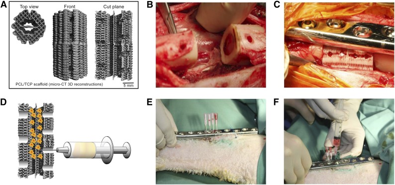
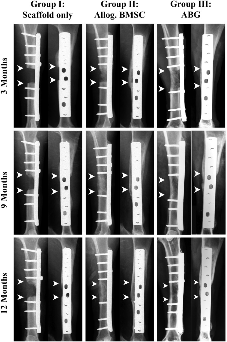
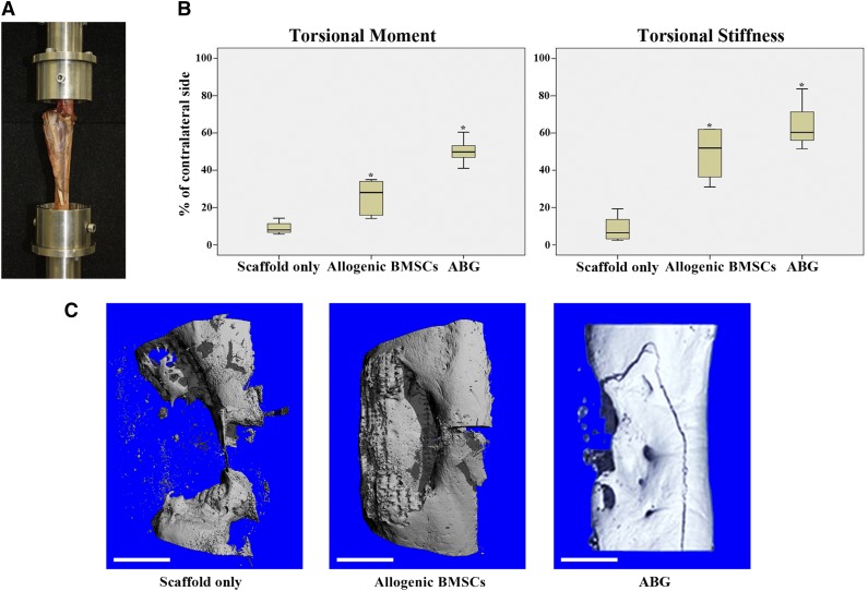
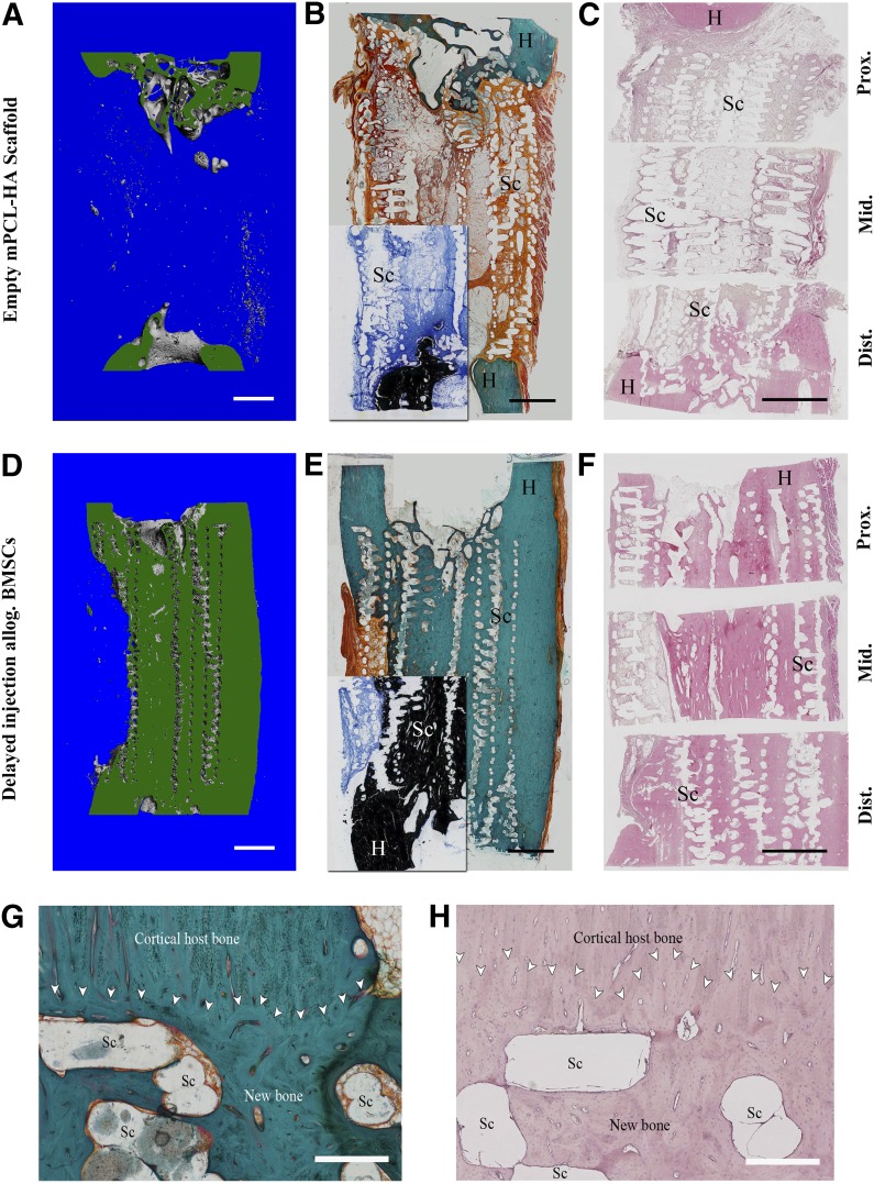
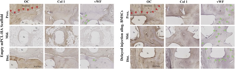
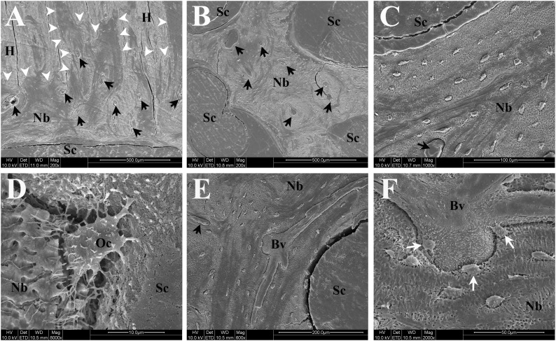
Cell-based tissue engineering approaches are promising strategies in the field of regenerative medicine. However, the mode of cell delivery is still a concern and needs to be significantly improved. Scaffolds and/or matrices loaded with cells are often transplanted into a bone defect immediately after the defect has been created. At this point, the nutrient and oxygen supply is low and the inflammatory cascade is incited, thus creating a highly unfavorable microenvironment for transplanted cells to survive and participate in the regeneration process. We therefore developed a unique treatment concept using the delayed injection of allogenic bone marrow stromal cell (BMSC) sheets to regenerate a critical-sized tibial defect in sheep to study the effect of the cells' regeneration potential when introduced at a postinflammatory stage. Minimally invasive percutaneous injection of allogenic BMSCs into biodegradable composite scaffolds 4 weeks after the defect surgery led to significantly improved bone regeneration compared with preseeded scaffold/cell constructs and scaffold-only groups. Biomechanical testing and microcomputed tomography showed comparable results to the clinical reference standard (i.e., an autologous bone graft). To our knowledge, we are the first to show in a validated preclinical large animal model that delayed allogenic cell transplantation can provide applicable clinical treatment alternatives for challenging bone defects in the future.

Keywords: Allogenic; Bone regeneration; Bone tissue engineering; Cell injection; Large bone defect; Mesenchymal stem cells; Sheep.


Figures

Figure 1.
Figure 1.
Scaffold design and surgical procedure. (A): Micro-CT 3D reconstruction of PCL-hydroxyapatite scaffold. (B): Posterior three holes placed in proximity to neurovascular bundle. (C): Surgical implantation in situ with four holes for delayed injection of bone marrow stromal cells (BMSCs) placed next to dynamic compression (DC) plate. (D): Schematic illustration of injection of BMSCs into the scaffold. (E, F): Minimally invasive percutaneous delayed BMSC injection procedure using another DC plate as a template to localize the injection holes and four separate needles to inject the cells into the scaffold. Abbreviations: 3D, three-dimensional; CT, computed tomography; PCL, polycaprolactone; TCP, tricalcium phosphate.
Figure 2.
Figure 2.
Representative clinical radiographic images at 3, 9, and 12 months after surgery. Defect reconstructed with polycaprolactone-hydroxyapatite (PCL-HA) scaffolds only (group I), PCL-HA scaffolds seeded with allogenic BMSCs (group II), and defects reconstructed with application of ABG (group III). Arrowheads indicate margins of bone defect. The images show radiographic signs of profound new bone formation and bridging of the defect site in the BMSC group and ABG group. The scaffold-only group shows attenuated signs of new bone formation with no bridging of the defect site. Abbreviations: ABG, autologous bone graft; Allog., allogenic; BMSCs, bone marrow stromal cells.
Figure 3.
Figure 3.
Biomechanical testing and microcomputed tomography (micro-CT) analysis. (A): Biomechanical testing was performed with both ends of the tibia embedded in methylmethacrylate with the tibial axis vertically aligned. (B): Results of biomechanical testing after 12 months for maximal torsional moment and torsional stiffness. Box plots demonstrate median values with first and third quartile in all experimental groups. Error bars represent maximum and minimum values. Asterisks indicate statistical significance (p < .05). (C): Representative three-dimensional reconstructions of micro-CT scans (proximal bone end facing upward) for scaffold-only group, allogenic BMSC group, and ABG group. Fracture line visible resulted from biomechanical testing (torsion until failure) before micro-CT analysis. Scale bars = 1 cm (C). Abbreviations: ABG, autologous bone graft; BMSC, bone marrow stromal cell.
Figure 4.
Figure 4.
Microcomputed tomography analysis at 12 months after surgery. (A): Image illustrating three different areas of interest. (B): Total bone volume in complete defect area. (C): Bone volume results in different areas of interest. Box plot demonstrating median amounts of newly formed bone with first and third quartile within the 3-cm defects 12 month after surgery. Error bars represent maximum and minimum values. Abbreviations: ABG, autologous bone graft; Allog., allogenic; BMSC, bone marrow stromal cell.
Figure 5.
Figure 5.
Representative images of histological staining for the empty scaffold group (A–C) and the allogenic BMSC group (D–H). Corresponding longitudinal sections (in green) through the three-dimensional reconstruction of mineralized tissue within the defect from microcomputed tomography data (A, D) are shown for comparison. Goldner’s trichrome stain (B, E) showed significant amount of new bone formation in the allogenic BMSC group compared with the empty scaffold group. Von Kossa/McNeal stain (insets in [B] and [E]) of corresponding areas confirm formation of mineralized tissue. Hematoxylin and eosin staining (C, F) also showed formation of new bone attenuated on scaffold-only group. Scaffold appears as a void owing to dissolution of PCL by xylene during processing. (G, H): Detailed view of host bone-scaffold interface of representative sample from allogenic BMSC group; Goldner’s trichrome stain (G) and hematoxylin and eosin stain (H). Good osteointegration of the scaffold and good bonding of the newly formed bone tissue with the host bone is visible. Scaffold struts appear as void owing to dissolving of PCL-HA by xylene during preparation. White arrowheads indicate interface between new bone/scaffold and host bone. Scale bars = 5 mm (A–F) and 500 µm (G, H). Abbreviations: Allog., allogenic; BMSC, bone marrow stromal cell; Dist., distal; H, host bone; mPCL-HA, medical grade polycaprolactone-hydroxyapatite; Mid., middle; Prox., proximal; Sc, scaffold struts.
Figure 6.
Figure 6.
Representative images of immunohistochemical analysis using antibodies against OC, Col 1, and vWF. Red arrowheads indicate host bone-new bone/scaffold interface; green arrowheads indicate vasculature. Early osteogenic marker Col 1 was found in newly formed bone of all segments of the allogenic BMSC group and newly formed bone proximally and distally and in fibrous tissue in the defect middle for the scaffold-only group. Late osteogenic marker OC was found in tissue-engineered bone in all segments in the allogenic BMSC group but only at the proximal and distal host bone-scaffold interfaces in the scaffold-only group. Profound neovascularization of the new bone tissue in all segments was present in the allogenic BMSC group (green arrowheads indicate blood vessels stained positively for vWF in new bone). Scaffold-only group showed new blood vessels in the proximal and distal interface areas. Scale bars = 100 µm. Abbreviations: Allog., allogenic; BMSC, bone marrow stromal cell; Col 1, collagen 1; Dist., distal; Mid., middle; mPCL-HA, medical grade polycaprolactone-hydroxyapatite; OC, osteocalcin; Prox., proximal; Sc, scaffold struts; vWF, von Willebrand factor.
Figure 7.
Figure 7.
Representative scanning electron microscopic images of newly formed bone and interfaces with scaffold/host bone in the delayed injection bone marrow stromal cell group. Black arrows indicate blood vessels within newly formed bone. (A): Interface of newly formed bone with host bone and scaffold showing excellent osteointegration (white arrowheads indicate host bone-new bone interface). (B): Osteointegration of newly formed bone with scaffold in the middle of the defect. (C): Higher magnification image showing integration between scaffold strut and tissue-engineered bone. (D): High-resolution image of an osteocyte partly embedded in newly formed bone matrix and also directly attaching to rough surface of scaffold strut (calcium-phosphate coating). (E): Image of large mature blood vessel with multiple branches inside tissue-engineered bone. (F): High-resolution image of blood vessels shown in (E) depicting close proximity and interaction of osteocytes (white arrows) to/with blood vessel. Fissures visible in images are artifacts resulting from preparation process of specimen. Abbreviations: Bv, blood vessel; H, host bone; Nb, newly formed bone; Oc, osteocyte; Sc, scaffold struts.

References

    1. Xu XQ, Sun W. Perspective from the heart: the potential of human pluripotent stem cell-derived cardiomyocytes. J Cell Biochem. 2013;114:39–46. - PubMed
    1. Vono R, Spinetti G, Gubernator M, et al. What’s new in regenerative medicine: Split up of the mesenchymal stem cell family promises new hope for cardiovascular repair. J Cardiovasc Transl Res. 2012;5:689–699. - PubMed
    1. Domínguez-Bendala J, Lanzoni G, Inverardi L, et al. Concise review: Mesenchymal stem cells for diabetes. Stem Cells Translational Medicine. 2012;1:59–63. - PMC - PubMed
    1. Jackson WM, Nesti LJ, Tuan RS. Concise review: Clinical translation of wound healing therapies based on mesenchymal stem cells. Stem Cells Translational Medicine. 2012;1:44–50. - PMC - PubMed
    1. Bernardo ME, Fibbe WE. Safety and efficacy of mesenchymal stromal cell therapy in autoimmune disorders. Ann NY Acad Sci. 2012;1266:107–117. - PubMed

Publication types

LinkOut - more resources