Review
. 2015 Apr;95(2):549-601.
doi: 10.1152/physrev.00035.2013.

Regulation of increased blood flow (hyperemia) to muscles during exercise: a hierarchy of competing physiological needs

Affiliations
Review

Regulation of increased blood flow (hyperemia) to muscles during exercise: a hierarchy of competing physiological needs

Michael J Joyner et al. Physiol Rev. 2015 Apr.

Abstract

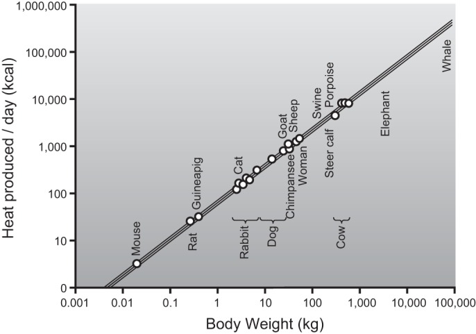
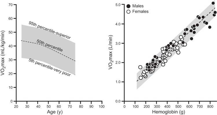
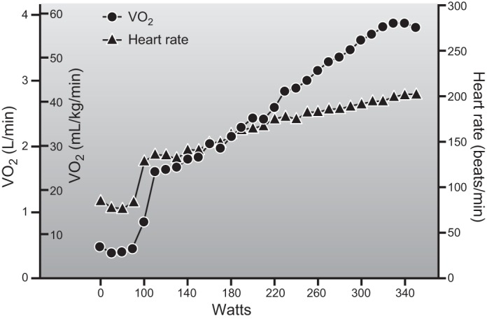
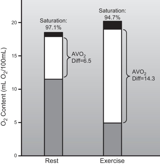
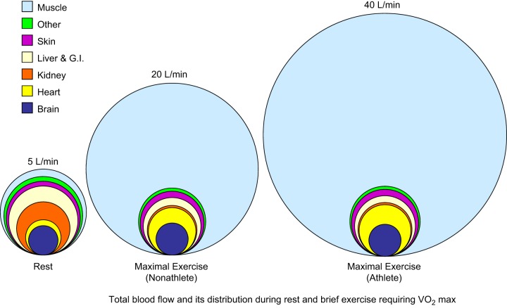
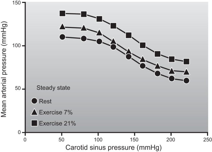
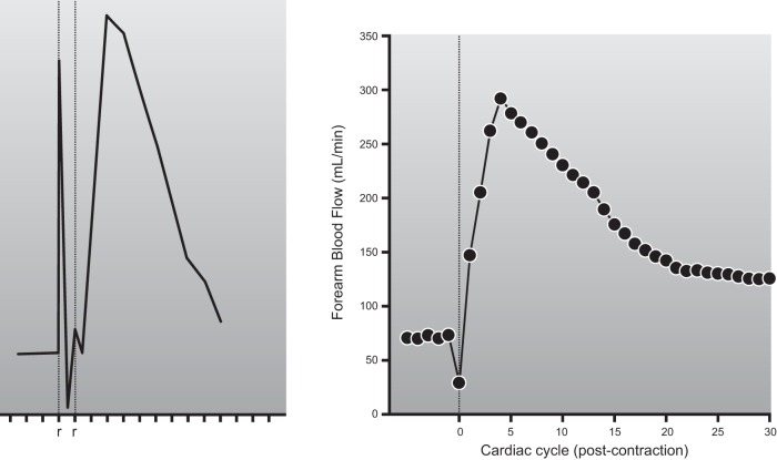
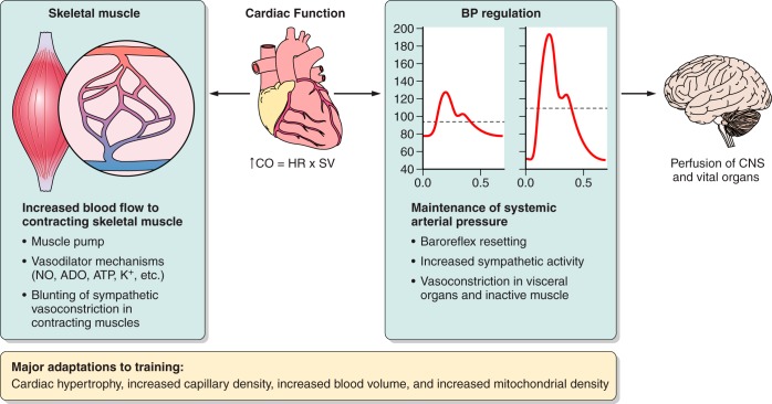
This review focuses on how blood flow to contracting skeletal muscles is regulated during exercise in humans. The idea is that blood flow to the contracting muscles links oxygen in the atmosphere with the contracting muscles where it is consumed. In this context, we take a top down approach and review the basics of oxygen consumption at rest and during exercise in humans, how these values change with training, and the systemic hemodynamic adaptations that support them. We highlight the very high muscle blood flow responses to exercise discovered in the 1980s. We also discuss the vasodilating factors in the contracting muscles responsible for these very high flows. Finally, the competition between demand for blood flow by contracting muscles and maximum systemic cardiac output is discussed as a potential challenge to blood pressure regulation during heavy large muscle mass or whole body exercise in humans. At this time, no one dominant dilator mechanism accounts for exercise hyperemia. Additionally, complex interactions between the sympathetic nervous system and the microcirculation facilitate high levels of systemic oxygen extraction and permit just enough sympathetic control of blood flow to contracting muscles to regulate blood pressure during large muscle mass exercise in humans.


Figures

Figure 1.
Figure 1.
Blood flow responses to single handgrip contractions of 0.33 s (left panel) and steady-state 1-leg kicking performed for a number of minutes (right panel). For both forms of exercise there is a linear relationship between exercise intensity and the blood flow response. This figure exemplifies the relationship between skeletal muscle metabolic demand and exercise hyperemia. The handgrip data also suggest that the rapid increases in blood flow to contracting muscles during exercise is due to vasodilation in the active muscles. The similarity of the responses in normal and sympathectomized limbs indicates that it is not dependent on sympathetic vasodilator nerves. For details, see Refs. and .
Figure 2.
Figure 2.
The relationship between body size on the x-axis and resting metabolic rate (heat production in kcal/day) on the y-axis. This figure shows that for every 10-fold (log) increase in body weight, the increase in resting metabolic rate is proportionally less than 1 log. This fundamental observation related to scaling helps explain why resting metabolic rate is lower on a per kilogram basis in larger animals. This type of analysis can also be used to compare exercise responses in elite human athletes of differing sizes. [Adapted from Kleiber (258).]
Figure 3.
Figure 3.
Population normal values for maximal oxygen consumption in ∼45,000 United States males expressed in ml·kg−1min−1 are shown in the left panel. For women, values and ranges 10–15% lower would typically be expected. The right panel demonstrates that total body hemoglobin is an important determinant of V̇o2max. This is because whole body hemoglobin content, along with maximum cardiac output, are major determinants of maximum oxygen delivery during exercise. This figure also shows the wide range of values for maximum oxygen uptake when considered in either relative (e.g., per kg; left panel) or absolute (l/min; right panel) terms. [Adapted from Joyner (238) and Trappe et al. (472).]
Figure 4.
Figure 4.
Individual record from a V̇o2max test in a well-trained but nonelite athletic male cyclist (18 yr of age) who weighed 67 kg. The x-axis is power output, and the y-axis is oxygen consumption both in l/min and scaled for body weight. There is a progressive increase in V̇o2 as power output increases with a leveling off at the highest work rates. This is accompanied by a linear increase in heart rate up to a value of ∼200 beats/min which is typical for a healthy young male. A V̇o2max value in the high 50s is typically attainable in young healthy lean male subjects who have participated in prolonged and intense exercise training. (Figure provided by Dr. Blair Johnson, unpublished observations.)
Figure 5.
Figure 5.
The classic data from Mitchell, Sproule, and Chapman, demonstrating oxygen-carrying capacity, arterial oxygen saturation, and mixed venous oxygen saturation in an untrained young healthy subject at rest and during maximal exercise. This figure emphasizes that while only ∼33% of the oxygen leaving the heart is extracted during rest, ∼70–75% can be extracted during heavy exercise in an untrained subject. The increase in whole body systemic oxygen extraction with exercise is facilitated by very high levels of extraction across the exercising muscle beds in conjunction with redistribution of blood flow from less active muscles and visceral organs. For details, see Ref. .
Figure 6.
Figure 6.
Schematic showing idealized distribution of blood flow at rest (left) and during maximum exercise in a healthy untrained young male subject (middle) and an elite endurance athlete (right). At rest, cardiac output is ∼5 l/min and rises to ∼20 l/min in the untrained subject during maximum exercise. Values of ∼40 l/min have been reported in elite endurance athletes. As cardiac output rises with exercise, brain blood flow remains constant (or increases slightly) while blood flow to the heart increases to meet the increased demands for myocardial blood flow that are primarily associated with exercise-induced increases in heart rate. Skeletal muscle blood flow increases dramatically, while blood flow to other tissues, especially the abdominal viscera and kidneys, is reduced. During heavy exercise, the vast increase in cardiac output is directed almost exclusively to contracting skeletal and cardiac muscles. Since maximum heart rate is similar in both young healthy subjects and elite athletes, the primary factor responsible for the very high cardiac outputs in the elite athlete is an extremely high stroke volume facilitated by a large compliant left ventricle. Ideas synthesized from Refs. , , , , , , –, , .
Figure 7.
Figure 7.
Transformational findings from three of the landmark studies showing that blood flow to contracting skeletal muscles during exercise could be much higher than previously imagined. The left panel is data from rats during treadmill running, the middle panel is from dogs, and the right panel is from humans during 1-leg kicking. The rat data show blood flow responses in ml·min−1·100 g−1 in various compartments of the hindlimb as running speed increased (VI, vastus intermedius; VLR, VLM, VLW, red, middle, and white vastus lateralis, respectively). The dog data show hindlimb blood flow responses vs. percent of V̇o2max generated during treadmill running. The human data show the blood flow responses in l/min vs. power output during 1-leg kicking at a rate of 60 kicks/min with the quadriceps (2–3 kg). The rat and dog data were obtained using radiolabeled microspheres and the human data via thermodilution. [Adapted from Andersen and Saltin (6), Armstrong and Laughlin (15), and Musch et al. (333).]
Figure 8.
Figure 8.
The distribution of blood flow to the arms and legs in a hypothetical elite cross-country skier during heavy exercise. The left panel shows that legs receive ∼20 l/min of blood flow during maximal diagonal skiing with only modest arm effort. Under these circumstances, cardiac output exceeds 30 l/min and mean arterial pressure (MAP) is ∼95 mmHg. The right panel shows what happens if the maximum values for double arm poling (DP) are added to the maximum values for the legs from diagonal skiing. If both beds dilated “maximally” and cardiac output remained constant, mean arterial pressure would fall to an estimated 75 mmHg. Under these circumstances, an additional ∼4 l/min of cardiac output would need to be generated to maintain mean arterial pressure at 95 mmHg. One implication of this figure is that at least some vasoconstriction in the contracting muscles is required to maintain arterial pressure during heavy exercise even in elite athletes who possess very high values for maximum cardiac output. [Adapted from Calbet et al. (67).]
Figure 9.
Figure 9.
Blood pressure responses to supine and head-down tilt exercise in an individual with autonomic failure as a result of surgical sympathectomy of the thoracolumbar sympathetic chain. The fall in blood pressure during supine exercise highlights the need for the sympathetic nervous system to restrain blood flow to contracting skeletal muscles for the purposes of regulating blood pressure. The fact that this fall in blood pressure also occurred when venous return was maximized by 15% head-down tilt emphasizes this point. The x-axis in the figures represents time with each vertical line representing 10 s. For details, see Ref. .
Figure 10.
Figure 10.
Demonstration in chronically instrumented dogs of baroreceptor resetting during exercise. This record was generated in animals that had undergone isolation of the carotid sinuses, permitting pressure in the carotid sinus to be controlled independently of arterial pressure. The input was carotid sinus pressure (x-axis); the output was systemic pressure measured in the whole animal (y-axis). During exercise, baroreceptor regulation of heart rate was reset to defend a higher arterial pressure, but the stimulus response curve to a given change in pressure was similar. Exercise was performed while running on a treadmill at 5.5 km/h up either a 7 or 21% grade. [Adapted from Joyner (237).]
Figure 11.
Figure 11.
The left panel is one of the original qualitative (no units provided) records of skeletal muscle blood flow (measured by timed collection of venous outflow) from the hindlimb of a dog whose skeletal muscle was stimulated to contract. Each tick mark on the x-axis represents a time interval of 5 s. The right panel shows a quantitative (ml/min) record of the brachial artery blood flow response to a single brief forearm contraction in a human more than 130 yr later. The key point is that with contraction there is an increase in venous effluent as blood is expelled from the muscle and veins. Arterial inflow stops or is attenuated as the contracting muscles compress the microcirculation of the skeletal muscle. With release of the electrical stimulation or voluntary handgrip contraction, there is a large increase in flow that declines rapidly. As is the case with the left panel in Figure 1, the marked and almost immediate rise in blood flow after the contraction or handgrip stops suggests that rapid vasodilation in the contracting skeletal muscles is a key determinant of exercise hyperemia. For details related to the left panel, see Ref. . Figure in the right panel are unpublished observations from our lab.
Figure 12.
Figure 12.
An individual record (compressed) demonstrating recovery of brachial artery blood velocity during acute reductions in perfusion pressure via intra-arterial balloon inflation during rhythmic handgripping at 20% maximal voluntary contraction. Exercise caused a rapid increase in blood velocity, while balloon inflation caused a fall in blood velocity that recovered within a couple of minutes. Breaks in velocity signal indicate times of image acquisition for diameter measurements, and breaks in arterial pressure tracing indicate times for blood sampling.
Figure 13.
Figure 13.
Forearm blood flow responses to mental stress. The left panel is an individual record showing the responses to severe mental stress as outlined in the text and comes from the classic study of Blair et al. (46). Note that the rise in flow is not immediate and is attenuated by brachial artery administration of atropine. The right panel shows the forearm blood flow responses in a more controlled form of mental stress (Stroop color word test); the open circles show the control forearm, and the closed circles show the forearm treated with a brachial artery infusion of the nitric oxide synthase (NOS) inhibitor l-NMMA. When the l-NMMA was allowed to washout for an hour, the responses were similar in both forearms. This demonstrates that the vasodilator responses to mental stress can be eliminated in large part by administration of the NOS inhibitor l-NMMA. For many years it was assumed that this response was due primarily to activation of vasodilating sympathetic cholinergic nerves. However, anatomical studies have failed to reveal the existence of such nerves in humans, and the current explanation for this rise in flow centers on vasodilation evoked via activation of β2-adrenergic receptors and mechanical stimulation of the vascular endothelium. Both mechanisms have a likely NO component. [Adapted from Blair et al. (46) and Dietz et al. (126).]
Figure 14.
Figure 14.
Skeletal muscle blood flow responses to auto-perfusion generated when a circuit connecting the hindlimbs of anesthetized pigs is isolated from the heart and the hindlimbs are electrically stimulated to contract. The pumping actions of the muscle contractions can increase blood flow in the absence of the heart and a normal arterial pressure. This shows that the skeletal muscle pump operating in isolation can generate some blood flow, but this increase in flow is modest at best compared with the large increases in skeletal muscle blood flow associated with exercise, but interpretation is not completely straightforward in the absence of normal perfusion pressure and gas exchange. The solid lines with various symbols represent the individual responses from each of the five pigs studied. The dashed line represents the averaged response for the five pigs (mean is based on 1 experiment from each of the 5 pigs). [Adapted from Sheriff and Van Bibber (437).]
Figure 15.
Figure 15.
The effects of rhythmic (1 s inflation/2 s deflation) external cuff compressions on forearm blood flow. The rhythmic inflation of an external cuff around the forearm was designed to mimic the mechanical effects of contraction. It causes a modest increase in forearm blood flow when the arm is below heart level. In contrast, when the arm is at heart level, there is little increase in blood flow. In either case, the increase in flow associated with cuff compression is very modest compared with what can be achieved with either a single contraction or more prolonged rhythmic hand gripping. This figure also shows that the effects of the muscle pump are dependent on limb position and also provides evidence that the muscle pump acting alone can generate only modest levels of blood flow. [Adapted from Tschakovsky et al. (478).]
Figure 16.
Figure 16.
The vasodilator response to a single forearm contraction at 20% of maximum is reduced by ∼80% when nitric oxide, prostaglandin, and K+-mediated hyperpolarization vasodilator pathways are inhibited. Blockade of NO and PGs alone can reduce this response ∼50%, and blockade of K+-mediated hyperpolarization alone can reduce it by ∼60%. At this time, it is not known if blockade of all of these pathways in combination will affect the steady-state blood flow responses to exercise. This is perhaps the most complete blunting of a vasodilator response to contractions currently available and highlights the idea that the vasodilator responses to contractions are highly redundant. It is also interesting to note that the immediate vasodilator responses to contractions can be blunted in conditions like aging (76, 85, 253). (Figure provided by Drs. Anne Crecelius and Frank Dinenno.)
Figure 17.
Figure 17.
Effects of prolonged adenosine infusion on blood flow on an isolated dog hindlimb preparation. Prior to adenosine infusion, electrically evoked skeletal muscle contraction caused an increase in blood flow. Adenosine was then infused for 150 min, and the vasodilator response to adenosine waned over that time. After the dilator response waned, the increase in blood flow to contractions was still present. The tachyphylaxis to adenosine indicated that skeletal muscle blood vessels had become desensitized to its vasodilator effects. The continued vasodilation with contractions after vasodilator responsiveness to adenosine had waned indicates that adenosine is not obligatory to generate a normal vasodilator response to contractions. [Adapted from Hester et al. (210).]
Figure 18.
Figure 18.
Vasodilator responses in coronary circulation of dogs during exercise, under control conditions, and after triple blockade when nitric oxide, adenosine, and calcium ATP channels are all blocked using various pharmacological antagonists. Note that the rise in coronary blood flow in response to an increase in myocardial oxygen consumption generated by graded treadmill exercise is similar in both conditions. This is an example of vasodilator redundancy in the coronary circulation that is similar to the redundancy seen in many human and animal studies on the regulation of blood flow in contracting human and animal muscles. It is seen in both isolated preparations and during exercise in conscious animals. While there are key differences between the coronary and skeletal muscle circulations, the concept that redundant vasodilator mechanisms govern the blood flow responses to exercise is a key commonality, as are the many putative dilator substances and mechanisms that might be involved. [Adapted from Tune et al. (481).]
Figure 19.
Figure 19.
Individual record showing the critical importance of time resolution and redundant vasodilator pathways in evaluating blood flow responses to pharmacological blockade in contracting skeletal muscles. In this forearm handgripping study (rhythmic contractions performed at 10% of maximal voluntary contraction for 20 min), addition of the KATP channel blocker glibenclamide after combined blockade of nitric oxide and prostaglandin synthesis (l-NAME + Ketorolac) caused an impressive but temporary reduction in blood flow to contracting forearm muscles. This was followed by a compensatory hyperemic response and return to steady-state values. One possible interpretation is that the blood flow response to contractions became more dependent on KATP channels during blockade of nitric oxide and prostaglandin synthesis. Thus it fell dramatically when glibenclamide was administered. This fall in flow then caused a mismatch between oxygen delivery and metabolism in the contracting muscles that evoked adenosine release. This caused a brief hyperemic response above the normal blood flow value associated with contractions and then a return toward the steady-state value seen before any intervention. These observations emphasize the need for rapid time course measurements of flow (beat-to-beat) when pharmacological blocking agents were given during contractions. Otherwise, it might be possible to miss effects of the drug on the pathway of interest. The individual responses seen in the larger experiment were variable, highlighting the possibility that there might be significant individual variability in the pathways normally recruited to evoke exercise hyperemia. [Adapted from Schrage et al. (415).]
Figure 20.
Figure 20.
Blood flow responses to temporary reductions in blood flow and perfusion pressure to the rhythmically contracting forearm muscles (20 contractions/min at 20% of maximum) evoked by inflation of a balloon in the brachial artery. Under control conditions, downstream vasodilation as contractions continued caused a complete recovery of blood flow over 2–3 min. This recovery of flow was delayed and blunted by NOS inhibition with l-NMMA and further blunted by the adenosine antagonist aminophylline. While adenosine does not appear obligatory in the vasodilator responses to contractions under many circumstances, its role becomes more critical when blood flow to the contracting muscles is restricted.
Figure 21.
Figure 21.
The effects of sympathetic stimulation (carotid occlusion) on perfusion pressure (MBP) in an isolated hindlimb preparation perfused with a roller pump at different rates of flow. As flow increases at rest, there is a rise in pressure in the roller pump circuit that increases further when sympathetic activity is increased by activation of carotid baroreflexes. This observation is consistent with the idea that the skeletal muscle vessels are being vasoconstricted. During contractions, pressure in the circuit is lower and rises less as flow is increased as a result of vasodilation in the muscles. Additionally, sympathetic stimulation has no effect on perfusion pressure consistent with the idea that the sympathetic nerves' ability to evoke vasoconstriction is blunted in contracting skeletal muscles. This effect has been termed “functional sympatholysis,” a controversial term as discussed further in the text. While this figure shows a complete blunting of the sympathetic constrictor responses during contractions, the results were variable, and in some animals, the constrictor responses were not fully abolished (Mitchell, personal communication). For details, see Ref. .
Figure 22.
Figure 22.
The effects of tyramine infusion on brachial artery vasodilator responses at rest and during rhythmic forearm exercise (20 contractions/min for 8 min). Tyramine is the prototypical indirectly acting sympathomimetic amine that causes release of norepinephrine from the sympathetic nerves. At rest and during both moderate (6.4 kg) and heavy exercise (12.1 kg), increasing doses of tyramine (2, 4, and 8 μg·dl forearm volume−1·min−1) caused dose-dependent vasoconstriction. This constriction was blunted by moderate exercise and attenuated more during heavy exercise. The tyramine infusions were adjusted to account for differences in blood flow to ensure a similar concentration under each condition. Importantly, when brachial artery flow was raised with either sodium nitroprusside or adenosine in the absence of exercise, there was no blunting of the constrictor responses (data not shown). The results highlighted in this figure show that contractions blunt sympathetic vasoconstriction in humans but that some constrictor tone is still present. This tone is critical to restrain blood flow to the contracting muscles for the purposes of blood pressure regulation during large muscle mass or whole body exercise. [Adapted from Tschakovsky et al. (479).]
Figure 23.
Figure 23.
The effects of sympathetic nerve activation (SNA) on the diameter of a feed artery (FA) and 3A arteriole in a hamster skeletal muscle. Measurements were made at rest and during contractions. A shows the FA response. During rest, sympathetic simulation caused constriction. During contractions, the diameter of the FA increased by 50%, but this dilation was reversed and essentially eliminated by sympathetic stimulation. In contrast, B shows the response in a much smaller 3A arteriole. The impressive constriction seen during sympathetic stimulation at rest is followed by a doubling of diameter during contractions. This dilation is unaffected when sympathetic stimulation is superimposed. This figure illustrates that sympatholysis is most pronounced in the smallest resistance vessels closest to the capillaries, while the larger arterial elements of the skeletal muscle microcirculation remain subject to sympathetic vasoconstriction. If sympathetic vasoconstriction eliminates relatively upstream vasodilation in contracting muscles, this would restrain total muscle blood flow. However, sympatholysis in the smallest vessels might serve to optimize the distribution of flow towards the most metabolically active or stressed elements of the contracting muscle(s). The overall effects of such responses would be seen at the systemic level where such sympathetic restraint is critical to regulate mean arterial pressure and also explains the almost total extraction of oxygen across exercising skeletal muscle vascular beds under some circumstances including heavy large muscle mass or whole body exercise. [Adapted from Van Teeffelen and Segal (487).]
Figure 24.
Figure 24.
Summary figure of the relationships between the local factors causing blood flow to rise in contracting skeletal muscles, cardiac output, and the need to regulate arterial blood pressure to ensure the perfusion of the central nervous system (CNS) and other vital organs. As emphasized throughout this review, factors released by the contracting muscles act locally to evoke vasodilation and blunt sympathetic vasoconstriction (functional sympatholysis). These events require an increase in cardiac output that is also facilitated by the systemic actions of the muscle pump to augment venous return. At the same time, it operates to increase perfusion pressure and amplify the effects of the vasodilating substances in the skeletal muscles. The cardiac hypertrophy and increases in blood volume caused by training also permit higher levels of muscle blood flow in the trained state. All of these acute and chronic adaptions are balanced by the autonomic nervous system in a way the permits arterial blood pressure to be maintained.

Similar articles

Cited by

References

    1. Abrahams VC, Hilton SM. The role of active muscle vasodilatation in the alerting stage of the defence reaction. J Physiol 171: 189–202, 1964. - PMC - PubMed
    1. Alam M, Smirk FH. Observations in man upon a blood pressure raising reflex arising from the voluntary muscles. J Physiol 89: 372–383, 1937. - PMC - PubMed
    1. Amann M. Pulmonary system limitations to endurance exercise performance in humans. Exp Physiol 97: 311–318, 2012. - PMC - PubMed
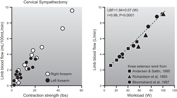
    1. Andersen P, Adams RP, Sjogaard G, Thorboe A, Saltin B. Dynamic knee extension as model for study of isolated exercising muscle in humans. J Appl Physiol 59: 1647–1653, 1985. - PubMed
    1. Andersen P, Henriksson J. Capillary supply of the quadriceps femoris muscle of man: adaptive response to exercise. J Physiol 270: 677–690, 1977. - PMC - PubMed

Publication types