Gastritis cystica profunda in a previously unoperated stomach: a case report
- PMID: 25834348
- PMCID: PMC4375605
- DOI: 10.3748/wjg.v21.i12.3759
Gastritis cystica profunda in a previously unoperated stomach: a case report
Abstract
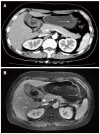
Gastritis cystica profunda is a relatively rare disease, usually observed at anastomotic sites in stomachs of patients that have undergone gastric procedures. We present the rare case of an elevated lesion in the anterior wall of the gastric antrum of a 43-year-old Chinese woman who had never undergone gastric surgery and had no gastrointestinal tract symptoms. Although the physical examination and laboratory data showed no abnormalities, endoscopic ultrasonography revealed an anechoic cystic structure. Abdominal computed tomography and magnetic resonance imaging showed the gastric wall of the greater curvature of the antrum was markedly and irregularly thickened, and mild to moderate enhancement was observed around the lesion with no enhancement in the central portion, suggestive of a gastrointestinal stromal tumor. The patient underwent a distal gastric resection of the 2.5 cm × 1.5 cm lesion. A postoperative pathologic examination showed dilated cystic glands in the muscularis mucosa and submucosal layers and erosion of the mucosal surface of the tumor, confirming the diagnosis of gastritis cystica profunda without malignancy.
Keywords: Gastritis cystica profunda; Hyperplastic polyp; Stomach.
Figures


References
-
- Littler ER, Gleibermann E. Gastritis cystica polyposa. (Gastric mucosal prolapse at gastroenterostomy site, with cystic and infiltrative epithelial hyperplasia) Cancer. 1972;29:205–209. - PubMed
-
- Xu G, Qian J, Ren G, Shan G, Wu Y, Ruan L. A case of gastritis cystica profunda. Ir J Med Sci. 2011;180:929–930. - PubMed
-
- Kurland J, DuBois S, Behling C, Savides T. Severe upper-GI bleed caused by gastritis cystica profunda. Gastrointest Endosc. 2006;63:716–717. - PubMed
-
- Matsumoto T, Wada M, Imai Y, Inokuma T. A rare cause of gastric outlet obstruction: gastritis cystica profunda accompanied by adenocarcinoma. Endoscopy. 2012;44 Suppl 2 UCTN:E138–E139. - PubMed
Publication types
MeSH terms
LinkOut - more resources
Full Text Sources
Other Literature Sources
Medical

