

Electrospun gelatin/polycaprolactone nanofibrous membranes combined with a coculture of bone marrow stromal cells and chondrocytes for cartilage engineering
- PMID: 25834428
- PMCID: PMC4370944
- DOI: 10.2147/IJN.S79461
Electrospun gelatin/polycaprolactone nanofibrous membranes combined with a coculture of bone marrow stromal cells and chondrocytes for cartilage engineering
Abstract
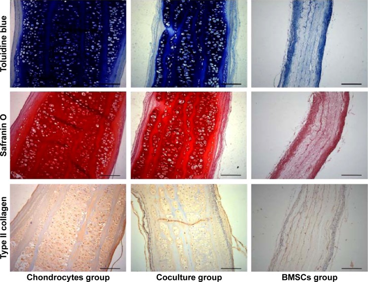
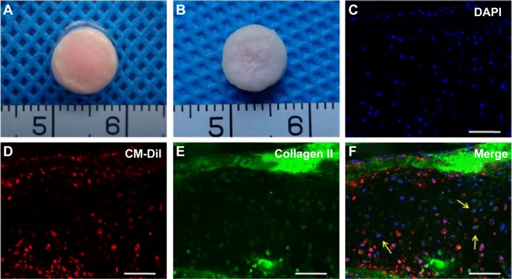
Electrospinning has recently received considerable attention, showing notable potential as a novel method of scaffold fabrication for cartilage engineering. The aim of this study was to use a coculture strategy of chondrocytes combined with electrospun gelatin/polycaprolactone (GT/PCL) membranes, instead of pure chondrocytes, to evaluate the formation of cartilaginous tissue. We prepared the GT/PCL membranes, seeded bone marrow stromal cell (BMSC)/chondrocyte cocultures (75% BMSCs and 25% chondrocytes) in a sandwich model in vitro, and then implanted the constructs subcutaneously into nude mice for 12 weeks. Gross observation, histological and immunohistological evaluation, glycosaminoglycan analyses, Young's modulus measurement, and immunofluorescence staining were performed postimplantation. We found that the coculture group formed mature cartilage-like tissue, with no statistically significant difference from the chondrocyte group, and labeled BMSCs could differentiate into chondrocyte-like cells under the chondrogenic niche of chondrocytes. This entire strategy indicates that GT/PCL membranes are also a suitable scaffold for stem cell-based cartilage engineering and may provide a potentially clinically feasible approach for cartilage repairs.
Keywords: cartilage tissue engineering; electrospinning; nanocomposite; nanomaterials; stem cells.
Figures


Similar articles
-
Engineering ear-shaped cartilage using electrospun fibrous membranes of gelatin/polycaprolactone.Biomaterials. 2013 Apr;34(11):2624-31. doi: 10.1016/j.biomaterials.2012.12.011. Epub 2013 Jan 24. Biomaterials. 2013. PMID: 23352044
-
Cartilage progenitor cells combined with PHBV in cartilage tissue engineering.J Transl Med. 2019 Mar 29;17(1):104. doi: 10.1186/s12967-019-1855-x. J Transl Med. 2019. PMID: 30925884 Free PMC article.
-
Electrospun collagen-poly(L-lactic acid-co-ε-caprolactone) membranes for cartilage tissue engineering.Regen Med. 2013 Jul;8(4):425-36. doi: 10.2217/rme.13.29. Regen Med. 2013. PMID: 23826697
-
Tissue engineering: chondrocytes and cartilage.Arthritis Res. 2002;4 Suppl 3(Suppl 3):S63-8. doi: 10.1186/ar561. Epub 2002 May 9. Arthritis Res. 2002. PMID: 12110124 Free PMC article. Review.
-
Gelatin Microsphere for Cartilage Tissue Engineering: Current and Future Strategies.Polymers (Basel). 2020 Oct 19;12(10):2404. doi: 10.3390/polym12102404. Polymers (Basel). 2020. PMID: 33086577 Free PMC article. Review.
Cited by
-
[Construction of tissue engineered cartilage based on acellular cartilage extracellular matrix oriented scaffold and chondrocytes].Zhongguo Xiu Fu Chong Jian Wai Ke Za Zhi. 2018 Mar 15;32(3):291-297. doi: 10.7507/1002-1892.201710095. Zhongguo Xiu Fu Chong Jian Wai Ke Za Zhi. 2018. PMID: 29806277 Free PMC article. Chinese.
-
An ECM-Mimicking, Mesenchymal Stem Cell-Embedded Hybrid Scaffold for Bone Regeneration.Biomed Res Int. 2017;2017:8591073. doi: 10.1155/2017/8591073. Epub 2017 Nov 15. Biomed Res Int. 2017. PMID: 29270436 Free PMC article.
-
Argon and Argon-Oxygen Plasma Surface Modification of Gelatin Nanofibers for Tissue Engineering Applications.Membranes (Basel). 2021 Jan 2;11(1):31. doi: 10.3390/membranes11010031. Membranes (Basel). 2021. PMID: 33401681 Free PMC article.
-
Functionalized Electrospun Scaffold-Human-Muscle-Derived Stem Cell Construct Promotes In Vivo Neocartilage Formation.Polymers (Basel). 2022 Jun 19;14(12):2498. doi: 10.3390/polym14122498. Polymers (Basel). 2022. PMID: 35746068 Free PMC article.
-
Surface modification of decellularized trachea matrix with collagen and laser micropore technique to promote cartilage regeneration.Am J Transl Res. 2019 Sep 15;11(9):5390-5403. eCollection 2019. Am J Transl Res. 2019. PMID: 31632518 Free PMC article.
References
-
- Vacanti CA, Paige KT, Kim WS, Sakata J, Upton J, Vacanti JP. Experimental tracheal replacement using tissue-engineered cartilage. J Pediatr Surg. 1994;29(2):201–204. discussion 204–205. - PubMed
-
- Wu W, Cheng X, Zhao Y, Chen F, Feng X, Mao T. Tissue engineering of trachea-like cartilage grafts by using chondrocyte macroaggregate: experimental study in rabbits. Artif Organs. 2007;31(11):826–834. - PubMed
-
- Barnewitz D, Endres M, Kruger I, et al. Treatment of articular cartilage defects in horses with polymer-based cartilage tissue engineering grafts. Biomaterials. 2006;27(14):2882–2889. - PubMed
-
- Liu Y, Zhang L, Zhou G, et al. In vitro engineering of human ear-shaped cartilage assisted with CAD/CAM technology. Biomaterials. 2010;31(8):2176–2183. - PubMed
Publication types
MeSH terms
Substances
LinkOut - more resources
Full Text Sources
Miscellaneous
