. 2015 Mar;18(1):44-9.
doi: 10.4048/jbc.2015.18.1.44. Epub 2015 Mar 27.

Ultrasonography-guided surgical clip placement for tumor localization in patients undergoing neoadjuvant chemotherapy for breast cancer

Affiliations

Ultrasonography-guided surgical clip placement for tumor localization in patients undergoing neoadjuvant chemotherapy for breast cancer

Inyoung Youn et al. J Breast Cancer. 2015 Mar.

Abstract

Purpose: We investigated the feasibility of using surgical clips as markers for tumor localization and their effect on the imaging evaluation of treatment responses after neoadjuvant chemotherapy (NAC).

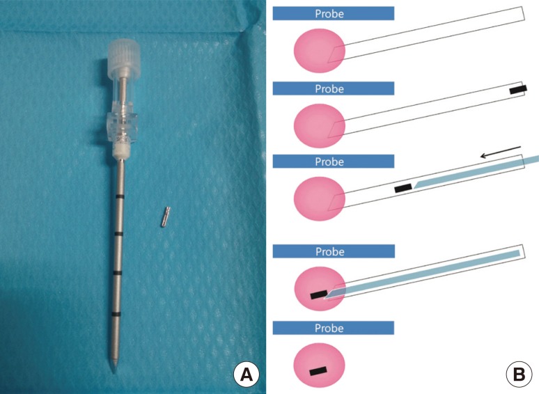
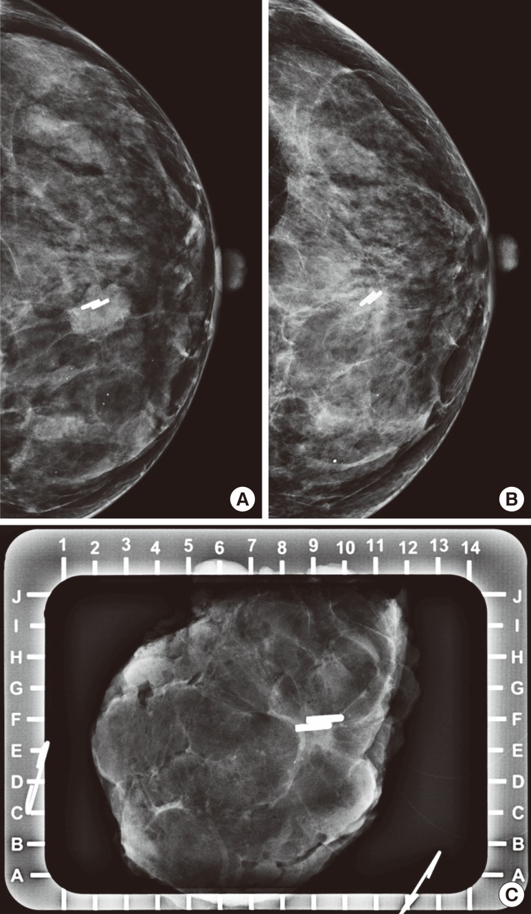
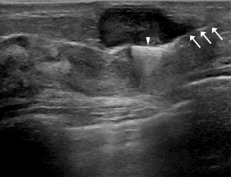
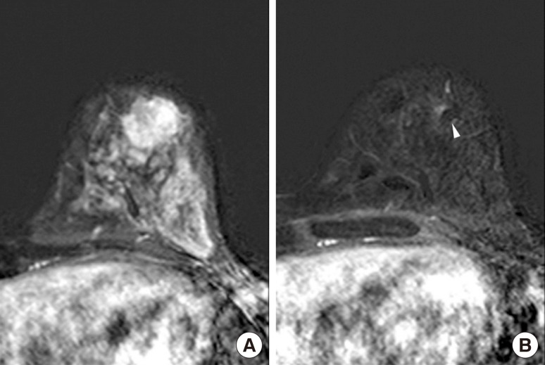
Methods: A total of 16 breast cancers confirmed by needle biopsy in 15 patients were included in this study from October 2012 to June 2014. Under ultrasonography (US)-guidance, the surgical clips were placed prior to NAC. Additional mammography, breast US, and breast magnetic resonance examinations were performed within 10 days before surgery. The time period from marker insertion to operation date was documented. Images acquired via the three modalities were evalu-ated for the following parameters: location of clip, clip migration (>1 cm), the presence of complications from clip placement, and the effect of clips on the assessment of treatment.

Results: The mean time period was 128.6±34.4 days (median, 132.0 days) from the date of clip insertion to the date of surgery. The mean number of inserted clips was 2.3±0.7 (median, 2.0). Clip migration was not visualized by imaging in any patient, and there were no complications reported. Surgical clips did not negatively affect the assessment of treatment responses to NAC.

Conclusion: Surgical clips may replace commercial tissue markers for tumor localization in breast cancer patients undergoing NAC without migration. Surgical clips are well tolerated and safe for the patient, easily visualized on imaging, do not interfere with treatment response, and are cost-effective.

Keywords: Breast neoplasms; Image-guided biopsy; Neoadjuvant therapy; Surgical instruments; Ultrasonography.


Conflict of interest statement

CONFLICT OF INTEREST: The authors declare that they have no competing interests.

Figures

Figure 1
Figure 1. Schematic diagram of the preoperative ultrasonography (US)-guided surgical clip insertion. (A) The coaxial guiding needle with an inner stylet and surgical clips. (B) Under US-guidance (blue), the coaxial guiding needle (white) is inserted into the the center of the breast cancer (pink), and one or two clips (black) are passed through. The inner stylet (light blue) is reinserted for pushing the clip.
Figure 2
Figure 2. Mammography of a 40-year-old woman who underwent ultrasonography-guided surgical clip insertion due to left breast cancer. (A) Postprocedural follow-up mammography was performed after clipping, and showed metal clips in the center of the proven malignant mass. (B) At preoperative final follow-up mammography, the clips were located in the proven malignant mass which had decreased in size. There was no evidence of clip migration or other complications. (C) Specimen mammography was performed immediately after surgery, and there were metal clips visualized in the proven malignant lesion without evidence of clip migration. The pathologic result was invasive carcinoma of no special type, and a clear tumor margin was observed.
Figure 3
Figure 3. Preoperative ultrasonography (US)-guided surgical clip insertion. On US images, the coaxial needle (arrows) is visible as an echogenic white line and the clips show a linear hyperechoic structure (arrowhead) in the center of the proven malignancy.
Figure 4
Figure 4. Magnetic resonance imaging (MRI) of a 40-year-old woman who underwent ultrasonography-guided surgical clip insertion due to left breast cancer. (A) There was a 19 mm-sized, fast, washout-enhancing malignancy at initial T1-weighted enhanced MRI with subtraction. (B) At preoperative follow-up MRI after neoadjuvant chemotherapy, a small signal void due to the clips (arrowhead) was observed in the center of the proven malignancy, which was much decreased in size and enhancement. However there was no difficulty in evaluating treatment response.

References

    1. Kaufmann M, von Minckwitz G, Bear HD, Buzdar A, McGale P, Bonnefoi H, et al. Recommendations from an international expert panel on the use of neoadjuvant (primary) systemic treatment of operable breast cancer: new perspectives 2006. Ann Oncol. 2007;18:1927–1934. - PubMed
    1. Oh JL, Nguyen G, Whitman GJ, Hunt KK, Yu TK, Woodward WA, et al. Placement of radiopaque clips for tumor localization in patients undergoing neoadjuvant chemotherapy and breast conservation therapy. Cancer. 2007;110:2420–2427. - PMC - PubMed
    1. Abdel-Razeq H, Marei L. Current neoadjuvant treatment options for HER2-positive breast cancer. Biologics. 2011;5:87–94. - PMC - PubMed
    1. Kim Z, Min SY, Yoon CS, Lee HJ, Lee JS, Youn HJ, et al. The basic facts of Korean breast cancer in 2011: results of a nationwide survey and breast cancer registry database. J Breast Cancer. 2014;17:99–106. - PMC - PubMed
    1. Lobbes MB, Prevos R, Smidt M, Tjan-Heijnen VC, van Goethem M, Schipper R, et al. The role of magnetic resonance imaging in assessing residual disease and pathologic complete response in breast cancer patients receiving neoadjuvant chemotherapy: a systematic review. Insights Imaging. 2013;4:163–175. - PMC - PubMed

LinkOut - more resources