Review
. 2015 Mar 31;7(4):2209-36.
doi: 10.3390/nu7042209.

Biomarkers of selenium status

Affiliations
Review

Biomarkers of selenium status

Gerald F Combs Jr. Nutrients. .

Abstract

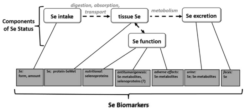
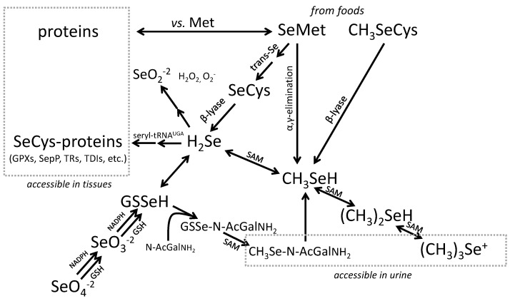
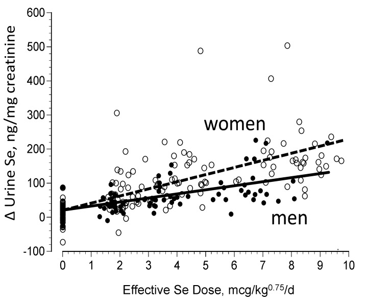
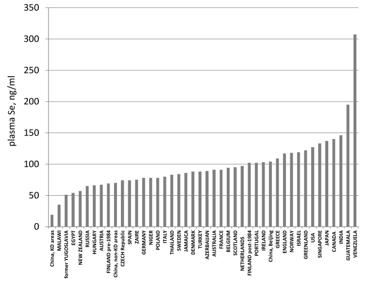
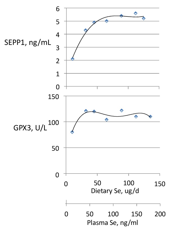
The essential trace element, selenium (Se), has multiple biological activities, which depend on the level of Se intake. Relatively low Se intakes determine the expression of selenoenzymes in which it serves as an essential constituent. Higher intakes have been shown to have anti-tumorigenic potential; and very high Se intakes can produce adverse effects. This hierarchy of biological activities calls for biomarkers informative at different levels of Se exposure. Some Se-biomarkers, such as the selenoproteins and particularly GPX3 and SEPP1, provide information about function directly and are of value in identifying nutritional Se deficiency and tracking responses of deficient individuals to Se-treatment. They are useful under conditions of Se intake within the range of regulated selenoprotein expression, e.g., for humans <55 μg/day and for animals <20 μg/kg diet. Other Se-biomarkers provide information indirectly through inferences based on Se levels of foods, tissues, urine or feces. They can indicate the likelihood of deficiency or adverse effects, but they do not provide direct evidence of either condition. Their value is in providing information about Se status over a wide range of Se intake, particularly from food forms. There is need for additional Se biomarkers particularly for assessing Se status in non-deficient individuals for whom the prospects of cancer risk reduction and adverse effects risk are the primary health considerations. This would include determining whether supranutritional intakes of Se may be required for maximal selenoprotein expression in immune surveillance cells. It would also include developing methods to determine low molecular weight Se-metabolites, i.e., selenoamino acids and methylated Se-metabolites, which to date have not been detectable in biological specimens. Recent analytical advances using tandem liquid chromatography-mass spectrometry suggest prospects for detecting these metabolites.


Figures

Figure 1
Figure 1
Selenium (Se) biomarkers and their relationships to the components of Se status.
Figure 2
Figure 2
Metabolism of Se. Note that inorganic Se can be incorporated into selenocystaine (SeCys)-containing selenoproteins by way of the obligate intermediate selenide (H2Se); while Se from selenomethionine (SeMet) can also be incorporated into the SeCys-containing proteins by way of catabolism to H2Se, and into other proteins directly by competing with methionine (Met) in protein synthesis. Other chemical symbols: SeO2−2 = selenium dioxide; SeO3−2 = selenite; SeO4−2 = selenate; CH3SeH = methyselenol; (CH3)2SeH = dimethylselenide’ (CH3)3Se+ = trimethyselenonium ion.
Figure 3
Figure 3
Changes in steady state urinary Se excretion in non-deficient adults after 13 months of supplementation with graded daily doses of SeMet [39].
Figure 4
Figure 4
Global variation in Se status as indicated by plasma Se concentration. Note: Because maximal selenoprotein expression is associated with plasma Se concentrations around 80 ng/mL, countries with averages less than ca. 100 ng/mL (parts of China, New Zealand, and much of Europe) are likely to have appreciable portions of their populations sub-optimally nourished with respect to Se. After Combs [23].
Figure 5
Figure 5
Effect of the form of dietary Se on tissue deposition of Se. After Reeves et al. [65].
Figure 5
Figure 5
Effect of the form of dietary Se on tissue deposition of Se. After Reeves et al. [65].
Figure 6
Figure 6
Stylized patterns of specific (solid areas, SeCys) and non-specific (hashed areas, presumed to be mostly SeMet in albumin) major components of plasma Se in subjects varying in Se status by consuming SeMet-containing foods. Not shown: Se occurring as SeMet in GPX3 and SEPP1, as their potential contributions to non-specific plasma Se are very small (<0.1%). Group labels: deficient = less than maximal selenprotein expression; adequate = maximal selenoprotein expression, i.e., meets nutritional requirement; “healthful” = supranutritional, including anti-tumorigenic doses; high = greater than needed for anti-tumorigenesis, but without adverse effects.
Figure 7
Figure 7
Relationship of plasma Se concentration and estimated Se intake in non-deficient Americans. (Regression: Sein, μg/kg0.75/day = 0.44 + 0.03 × Seplasma, ng/mL; dotted lines indicate 95% confidence limits) Recalculated from data of Combs et al. [50].
Figure 8
Figure 8
Relationships of GPX3 activity and SEPP1 concentration to dietary Se intake and plasma Se concentration in residents of a low-Se part of China. Subjects had an estimated average intake of 10 μg/day from dietary sources and had been supplemented with graded amounts of SeMet for 40 weeks. Estimated from the data of Xia et al. [91].

References

    1. Painter E.P. The chemistry and toxicity of selenium compounds, with special reference to the selenium problem. Chem. Rev. 1941;28:179–187.
    1. Schwarz K., Foltz C.M. Selenium as an integral part of factor 3 against dietary necrotic liver degeneration. J. Am. Chem. Soc. 1957;79:3292–3293. - PubMed
    1. Schwarz K., Bieri J.G., Briggs G.M., Scott M.L. Prevention of exudative diathesis in chicks by factor 3 and selenium. Proc. Soc. Exp. Biol. Med. 1957;95:621–629. doi: 10.3181/00379727-95-23308. - DOI - PubMed
    1. Patterson E.L., Milstrey R., Stokstad E.L.R. Effect of selenium in preventing exudative diathesis in chicks. Proc. Soc. Exp. Biol. Med. 1957;95:617–625. doi: 10.3181/00379727-95-23307. - DOI - PubMed
    1. Ge K., Xue A., Bai J., Wang S. Keshan Disease—An endemic cardiomyopathy in China. Virchow Arch. 1983;410:1–12. doi: 10.1007/BF00644785. - DOI - PubMed