. 2015 Apr 2;11(4):e1005065.
doi: 10.1371/journal.pgen.1005065. eCollection 2015 Apr.

Viable neuronopathic Gaucher disease model in Medaka (Oryzias latipes) displays axonal accumulation of alpha-synuclein

Affiliations

Viable neuronopathic Gaucher disease model in Medaka (Oryzias latipes) displays axonal accumulation of alpha-synuclein

Norihito Uemura et al. PLoS Genet. .

Abstract

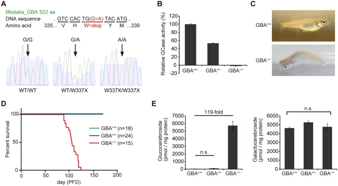
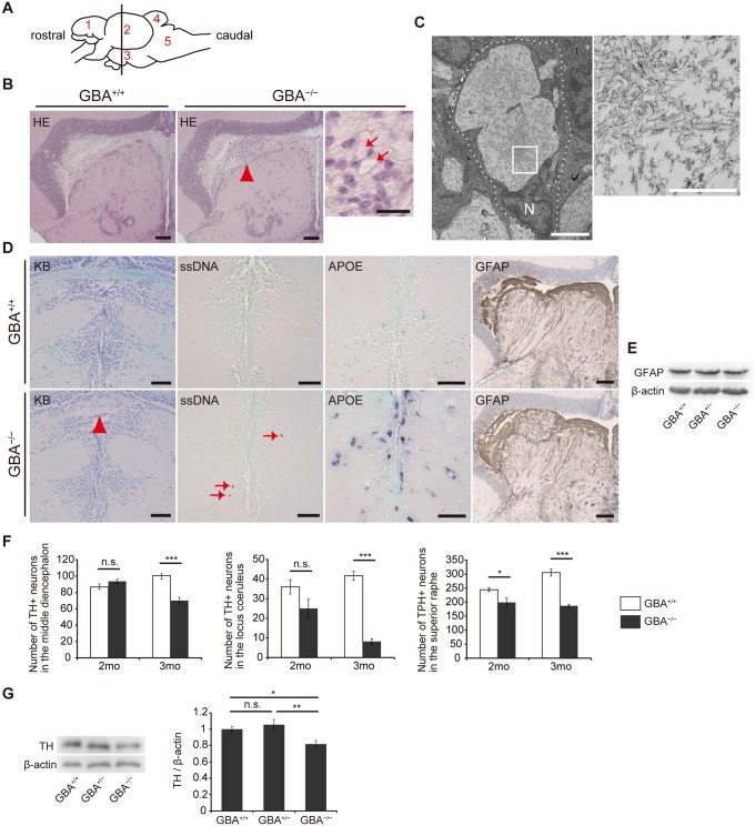
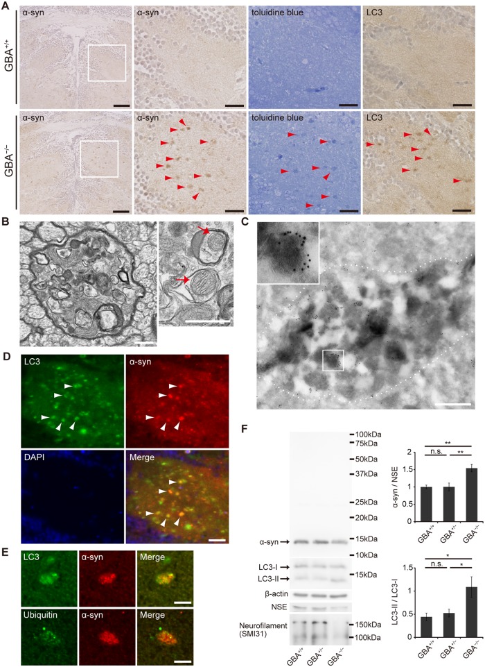
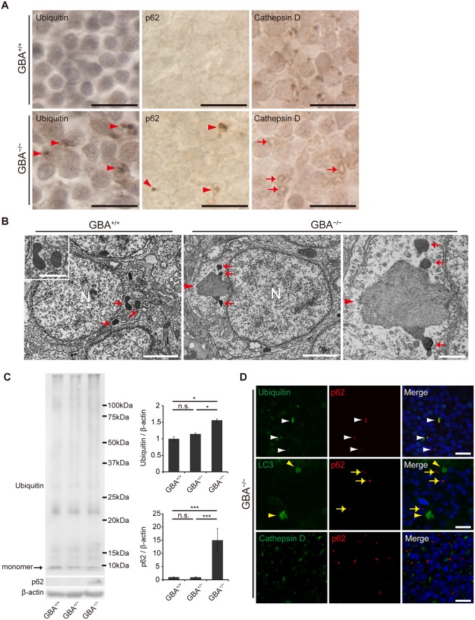
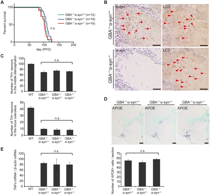
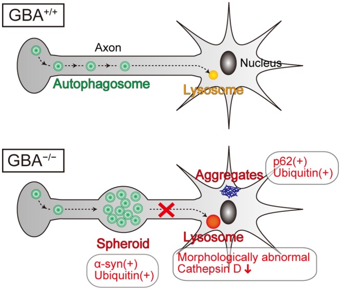
Homozygous mutations in the glucocerebrosidase (GBA) gene result in Gaucher disease (GD), the most common lysosomal storage disease. Recent genetic studies have revealed that GBA mutations confer a strong risk for sporadic Parkinson's disease (PD). To investigate how GBA mutations cause PD, we generated GBA nonsense mutant (GBA-/-) medaka that are completely deficient in glucocerebrosidase (GCase) activity. In contrast to the perinatal death in humans and mice lacking GCase activity, GBA-/- medaka survived for months, enabling analysis of the pathological progression. GBA-/- medaka displayed the pathological phenotypes resembling human neuronopathic GD including infiltration of Gaucher cell-like cells into the brains, progressive neuronal loss, and microgliosis. Detailed pathological findings represented lysosomal abnormalities in neurons and alpha-synuclein (α-syn) accumulation in axonal swellings containing autophagosomes. Unexpectedly, disruption of α-syn did not improve the life span, formation of axonal swellings, neuronal loss, or neuroinflammation in GBA-/- medaka. Taken together, the present study revealed GBA-/- medaka as a novel neuronopathic GD model, the pahological mechanisms of α-syn accumulation caused by GCase deficiency, and the minimal contribution of α-syn to the pathogenesis of neuronopathic GD.


Conflict of interest statement

The authors have declared that no competing interests exist.

Figures

Fig 1
Fig 1. Generation of GBA nonsense mutant medaka.
(A) Upper panel: DNA and amino acid sequences of GBA nonsense mutant medaka. Lower panels: Sequence data for each genotype. A = green, T = red, G = black, and C = blue. (B) Relative GCase activity in medaka brains (n = 10). (C) Abnormal posture (‘bent spine’) in GBA -/- medaka at 3 months. (D) Survival curves for each genotype. PFD, post-fertilization day. (E) Quantification of glucocerebroside and galactocerebroside in medaka brains at 3 months with SFC/MS/MS (n = 3–4). For all analyses, data are the mean ± standard error of the mean (SEM). n.s. means not significant.
Fig 2
Fig 2. Pathological analyses of GBA -/- medaka.
(A) Schematic of a lateral view of the medaka brain. Each number signifies a part of the brain. 1: telencephalon, 2: optic tectum, 3: diencephalon, 4: cerebellum, 5: medulla oblongata. The brain sections used for pathological analyses in the present study are illustrated by the vertical line. (B) Hematoxylin and eosin staining. Abnormal cells observed in the periventricular gray zone of the optic tectum (arrowhead) in GBA -/- medaka at 3 months. Scale bars, 50 μm. Right panel: High-magnification image showing abnormal cells with large vacuoles (arrows). Scale bar, 5 μm. (C) Transmission electron micrographs showing abnormal macrophage-like cells. Left panel: A whole-cell image of an abnormal macrophage-like cell. Dashed lines outline a whole abnormal cell. N, Nucleus. Scale bar, 2 μm. Right panel: High-magnification image of filamentous structures in vacuoles. Scale bar, 500 nm. (D) Klüver-Barrera (KB) staining, ssDNA immunohistochemistry, APOE in situ hybridization, and GFAP immunohistochemistry in the diencephalon. Commissura posterior with decreased Luxol fast blue staining intensity (arrowhead) and ssDNA-positive dead cells (arrows) in GBA -/- medaka. APOE in situ hybridization revealed proliferating activated microglia in GBA -/- medaka. The staining intensity and area of GFAP were not changed in GBA -/- medaka. Scale bars, 50 μm. (E) Western blot analysis of GFAP and β-actin. The expression level of GFAP was not changed among genotypes. (F) Number of TH-positive neurons in the middle diencephalon, number of TH-positive neurons in the locus coeruleus, and number of TPH-positive neurons in the superior raphe at 2 and 3 months. In GBA -/- medaka, progressive neuronal loss was observed in all types of neurons (n = 4–6, *p < 0.05, ***p < 0.001). (G) Western blot analysis of TH and β-actin (n = 7, *p < 0.05, **p < 0.01). For all analyses, data are the mean ± SEM.
Fig 3
Fig 3. Axonal swellings with α-syn accumulation in GBA -/- medaka.
(A) α-syn and LC3 immunohistochemistry, and toluidine blue staining in the diencephalon at 3 months. Left panels: α-syn accumulations were observed in GBA -/- medaka. Scale bars, 50 μm. Other panels: outlined area of left panels. The distribution of α-syn accumulations was similar to that of abnormal structures observed with toluidine blue staining and LC3 accumulations (arrowheads). Scale bars, 20 μm. (B) Transmission electron micrographs of axonal swellings in GBA -/- medaka. Left panel: a swelling of a myelinated axon containing vacuoles and electron-dense bodies. Right panel: vacuoles in an axonal swelling containing mitochondria (arrows). Scale bars, 500 nm. (C) Immunoelectron micrograph of an axonal swelling in a GBA -/- medaka with immunogold-labeled α-syn. Dashed lines outline an axonal swelling containing vacuoles and electron-dense bodies. The boxed area is enlarged in the inset. Scale bar, 500 nm. (D) Double immunostaining for LC3 (green) and α-syn (red) in GBA -/- medaka. Nuclei were visualized with DAPI (blue). α-syn accumulations colocalized with LC3 accumulations (arrowheads). Scale bar, 20 μm. (E) Conforcal microscope images of α-syn-positive axonal swellings. Upper panels: Double immunostaining for LC3 (green) and α-syn (red). A considerable portion of the α-syn signals colocalized with LC3 signals. Lower panels: Double immunostaining for ubiquitin (green) and α-syn (red). Ubiquitin colocalized with an α-syn accumulation. Scale bars, 5 μm. (F) Western blot analysis of α-syn, LC3, β-actin, NSE, and neurofilament at 3 months (n = 6–7, *p < 0.05, **p < 0.01). For all analyses, data are the mean ± SEM.
Fig 4
Fig 4. Impairment of the autophagy-lysosome pathway in GBA -/- medaka.
(A) Ubiquitin, p62, and Cathepsin D immunohistochemistry in the neuronal layer of the optic tectum at 3 months. Almost all of the cells in these figures are neurons. Ubiquitin-positive and p62-positive aggregates were observed in the perikarya of neurons in GBA -/- medaka at 3 months (arrowheads). Morphologically abnormal organelles with decreased Cathepsin D staining intensity were observed in GBA -/- medaka (arrows). Scale bars, 10 μm. (B) Transmission electron micrographs of neurons. Left panel: Neuron of a GBA +/+ medaka. Electron-dense organelles (arrows) are likely lysosomes. N, Nucleus. Scale bar, 2 μm. The inset shows a high-magnification image of electron-dense organelles. Scale bar, 500 nm. Middle panel: Neuron of a GBA -/- medaka. An aggregate containing filamentous structures (arrowhead) is continuous with an electron-dense organelle. N, Nucleus. Scale bar, 2 μm. Right panel: High-magnification image of an aggregate of filamentous structures (arrowhead) and electron-dense organelles (arrows) in a GBA -/- medaka. Scale bar, 500 nm. (C) Western blot analysis of ubiquitin, p62, and β-actin (n = 4–7, *p < 0.05, ***p < 0.001). (D) Conforcal microscope images of GBA -/- medaka. Upper panels: ubiquitin (green) and p62 (red). p62-positive aggregates colocalized with ubiquitin-positive aggregates (white arrowheads). Middle panels: LC3 (green) and p62 (red). p62-positive aggregates (yellow arrows) did not colocalize with LC3 accumulations (yellow arrowheads). Lower panels: Cathepsin D (green) and p62 (red). p62-positive aggregates did not colocalize with Cathepsin D-positive organelles. Nuclei were visualized with DAPI (blue). Scale bars, 10 μm. For all analyses, data are the mean ± SEM.
Fig 5
Fig 5. Disruption of α-syn did not change the pathological phenotypes in GBA -/- medaka.
(A) Survival curves for each genotype. Life spans were not changed among genotypes. (B) α-syn and LC3 immunohistochemistry in the diencephalon. α-syn accumulations were observed only in GBA -/- α-syn +/+ medaka (arrows). The number of LC3-positive puncta was not different between GBA -/- α-syn +/+ and GBA -/- α-syn -/- medaka (arrowheads). Scale bars, 20 μm. (C) Number of TH-positive neurons in the middle diencephalon and the locus coeruleus at 3 months (n = 6). (D) Upper panel: APOE in situ hybridization in the optic tectum and the diencephalon. Lower panel: The number of APOE-positive cells per section (n = 9). Scale bars, 50 μm. (E) Quantification of TNFα mRNA levels normalized to β-actin mRNA in medaka brains with quantitative RT-PCR (n = 8). For all analyses, data are the mean ± SEM.
Fig 6
Fig 6. Pathological findings and proposed pathological mechanisms in neurons of GBA -/- medaka.
Neurons of GBA -/- medaka showed lysosomal dysfunction, which is reflected in morphologically abnormal structures with decreased Cathepsin D staining intensity, and axonal swellings containing autophagosomes where α-syn and ubiquitin accumulate. p62-positive aggregates, which colocalize with ubiquitin, are not located in axonal swellings, but presumably in soma or dendrites. Considering the previous studies about the axonal transport of autophagosomes, we propose that GCase deficiency primarily causes lysosomal dysfunction, which leads to disrupted retrograde transport of autophagosomes in axons and formation of axonal swellings with α-syn accumulation.

Similar articles

Cited by

References

    1. Futerman AH, Zimran A (2006) Gaucher disease: CRC Press.
    1. Grabowski GA (2008) Phenotype, diagnosis, and treatment of Gaucher's disease. Lancet 372: 1263–1271. 10.1016/S0140-6736(08)61522-6 - DOI - PubMed
    1. Wong K, Sidransky E, Verma A, Mixon T, Sandberg GD, et al. (2004) Neuropathology provides clues to the pathophysiology of Gaucher disease. Mol Genet Metab 82: 192–207. - PubMed
    1. Eblan MJ, Goker-Alpan O, Sidransky E (2005) Perinatal lethal Gaucher disease: a distinct phenotype along the neuronopathic continuum. Fetal Pediatr Pathol 24: 205–222. - PubMed
    1. Neumann J, Bras J, Deas E, O'Sullivan SS, Parkkinen L, et al. (2009) Glucocerebrosidase mutations in clinical and pathologically proven Parkinson's disease. Brain 132: 1783–1794. 10.1093/brain/awp044 - DOI - PMC - PubMed

Publication types