

Thymosin beta 4 may translocate from the cytoplasm in to the nucleus in HepG2 cells following serum starvation. An ultrastructural study
- PMID: 25835495
- PMCID: PMC4383617
- DOI: 10.1371/journal.pone.0119642
Thymosin beta 4 may translocate from the cytoplasm in to the nucleus in HepG2 cells following serum starvation. An ultrastructural study
Abstract
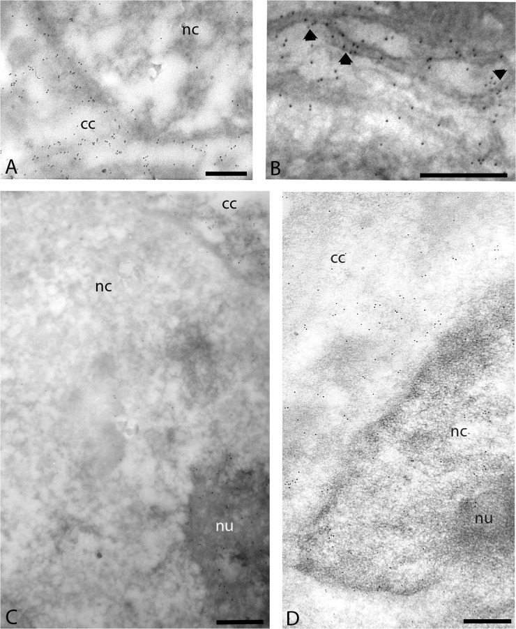
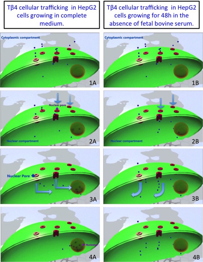
Due to its actin-sequestering properties, thymosin beta-4 (Tβ4) is considered to play a significant role in the cellular metabolism. Several physiological properties of Tβ4 have been reported;, however, many questions concerning its cellular function remain to be ascertained. To better understand the role of this small peptide we have analyzed by means of transmission immunoelectron microscopy techniques the ultrastructural localization of Tβ4 in HepG2 cells. Samples of HepG2 cells were fixed in a mixture of 3% formaldehyde and 0.1% glutaraldehyde in 0.1 M cacodylate buffer and processed for standard electron microscopic techniques. The samples were dehydrated in a cold graded methanol series and embedded in LR gold resin. Ultrathin sections were labeled with rabbit antibodies to Tβ4, followed by gold-labeled goat anti-rabbit, stained with uranyl acetate and bismuth subnitrate, observed and photographed in a JEOL 100S transmission electron microscope. High-resolution electron microscopy showed that Tβ4 was mainly restricted to the cytoplasm of HepG2 growing in complete medium. A strong Tβ4 reactivity was detected in the perinuclear region of the cytoplasmic compartment where gold particles appeared strictly associated to the nuclear membrane. In the nucleus specific Tβ4 labeling was observed in the nucleolus. The above electron microscopic results confirm and extend previous observations at light microscopic level, highlighting the subcellular distribution of Tβ4 in both cytoplasmic and nuclear compartments of HepG2 cells. The meaning of Tβ4 presence in the nucleolus is not on the best of our knowledge clarified yet. It could account for the interaction of Tβ4 with nucleolar actin and according with this hypothesis, Tβ4 could contribute together with the other nucleolar acting binding proteins to modulate the transcription activity of the RNA polymerases.
Conflict of interest statement
Figures
Similar articles
-
Cellular trafficking of thymosin beta-4 in HEPG2 cells following serum starvation.PLoS One. 2013 Aug 14;8(8):e67999. doi: 10.1371/journal.pone.0067999. eCollection 2013. PLoS One. 2013. PMID: 23967050 Free PMC article.
-
Ku80 as a novel receptor for thymosin beta4 that mediates its intracellular activity different from G-actin sequestering.J Biol Chem. 2008 Jan 18;283(3):1534-1544. doi: 10.1074/jbc.M707539200. Epub 2007 Nov 4. J Biol Chem. 2008. PMID: 17984099
-
Interleukin-18-mediated interferon-gamma secretion is regulated by thymosin beta 4 in human NK cells.Immunobiology. 2011 Oct;216(10):1155-62. doi: 10.1016/j.imbio.2011.04.002. Epub 2011 Apr 13. Immunobiology. 2011. PMID: 21742406
-
Platelet function and thymosin β4.Biol Chem. 2012 Jul;393(7):595-8. doi: 10.1515/hsz-2012-0131. Biol Chem. 2012. PMID: 22944663 Review.
-
Repolymerization of actin from actin:thymosin beta4 complex induced by diaphanous related formins and gelsolin.Ann N Y Acad Sci. 2010 Apr;1194:36-43. doi: 10.1111/j.1749-6632.2010.05467.x. Ann N Y Acad Sci. 2010. PMID: 20536448 Review.
Cited by
-
Effect of vitamin A deficiency on thymosin-β4 and CD4 concentrations.J Genet Eng Biotechnol. 2018 Jun;16(1):57-61. doi: 10.1016/j.jgeb.2017.10.007. Epub 2017 Oct 10. J Genet Eng Biotechnol. 2018. PMID: 30647705 Free PMC article.
-
The Hidden Function of Vitamin D.Open Access Maced J Med Sci. 2016 Dec 15;4(4):591-595. doi: 10.3889/oamjms.2016.134. Epub 2016 Nov 30. Open Access Maced J Med Sci. 2016. PMID: 28028396 Free PMC article.
-
Thymosin β4 cytoplasmic/nuclear translocation as a new marker of cellular stress. A Caco2 case study.RSC Adv. 2020 Mar 30;10(21):12680-12688. doi: 10.1039/c9ra10365a. eCollection 2020 Mar 24. RSC Adv. 2020. PMID: 35497634 Free PMC article.
-
Toward the renal vesicle: Ultrastructural investigation of the cap mesenchyme splitting process in the developing kidney.J Public Health Res. 2022 Oct 24;11(4):22799036221124076. doi: 10.1177/22799036221124076. eCollection 2022 Oct. J Public Health Res. 2022. PMID: 36310827 Free PMC article.
-
Thymosin β4 overexpression regulates neuron production and spatial distribution in the developing avian optic tectum.Histochem Cell Biol. 2017 May;147(5):555-564. doi: 10.1007/s00418-016-1529-1. Epub 2016 Dec 10. Histochem Cell Biol. 2017. PMID: 27942867
References
-
- Huff T, Müller CS, Otto AM, Netzker R, Hannappel E (2001) beta-Thymosins, small acidic peptides with multiple functions. Int J Biochem Cell Biol 33: 205–220. - PubMed
-
- Goldstein AL (2007) History of the discovery of the thymosins. Ann N Y Acad Sci 1112: 1–13. - PubMed
-
- Ballweber E, Hannappel E, Huff T, Stephan H, Haener M, Taschner N et al. (2002) Polymerisation of chemically cross-linked actin:thymosin beta(4) complex to filamentous actin: alteration in helical parameters and visualisation of thymosin beta(4) binding on F-actin. J Mol Biol 315: 613–625. - PubMed
-
- Goldstein AL, Hannappel E, Kleinman HK. (2005) Thymosin β4: actin-sequestering protein moonlights to repair injured tissues. Trends Mol Med 11: 421–429.6. - PubMed
Publication types
MeSH terms
Substances
LinkOut - more resources
Full Text Sources
Other Literature Sources
