

Relevance of the plasma membrane calcium-ATPase in the homeostasis of calcium in the fetal liver
- PMID: 25836032
- PMCID: PMC4594366
- DOI: 10.1080/15476278.2015.1011918
Relevance of the plasma membrane calcium-ATPase in the homeostasis of calcium in the fetal liver
Abstract
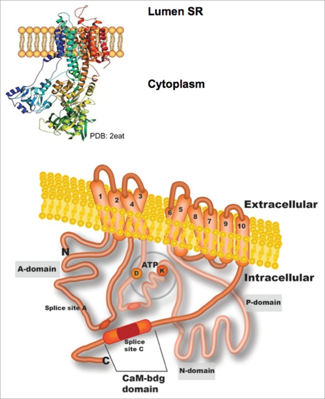
During the early stages of development, the embryo depends on the placenta as provider of oxygen and calcium, among other essential compounds. Although fetal liver accomplishes a well-known haematopoietic function, its contribution to calcium homeostasis upon development is poorly understood. The homeostasis of cell calcium contributes to diverse signaling pathways across developmental stages of most tissues and the calcium-ATPase located at the plasma membrane (PMCA) helps pumping excess calcium into the extracellular space. To date, the understanding of the equilibrium shift between PMCA isoforms during liver development is still missing. This review focuses on the characterization of the hepatic PMCA along the early stages of development, followed by a description of modern approaches to study calcium homeostasis involving several types of pluripotent cells. The application of interdisciplinary techniques to improve our understanding of liver development and the role calcium homeostasis plays in the definition of pathogenesis is also discussed.
Keywords: Ca2+ homeostasis; embryonic stem cells; fetal liver; induced pluripotent stem cells; plasma membrane calcium ATPase.
Figures
Similar articles
-
Is there a specific role for the plasma membrane Ca2+ -ATPase in the hepatocyte?Mol Cell Biochem. 2006 Apr;285(1-2):1-15. doi: 10.1007/s11010-005-9060-z. Epub 2006 Feb 14. Mol Cell Biochem. 2006. PMID: 16477375 Review.
-
Intracellular calcium homeostasis in Leishmania mexicana. Identification and characterization of a plasma membrane calmodulin-dependent Ca(2+)-ATPase.Biol Res. 1993;26(1-2):141-50. Biol Res. 1993. PMID: 7670527
-
Loss of calcium homeostasis leads to progressive phase of chlordecone-potentiated carbon tetrachloride hepatotoxicity.Toxicol Appl Pharmacol. 1993 Sep;122(1):77-87. doi: 10.1006/taap.1993.1174. Toxicol Appl Pharmacol. 1993. PMID: 7690997
-
A developmental profile of the levels of calcium pumps in chick cerebellum.J Neurochem. 2005 Nov;95(3):673-83. doi: 10.1111/j.1471-4159.2005.03401.x. Epub 2005 Aug 16. J Neurochem. 2005. PMID: 16104848
-
Plasma membrane Ca2+-ATPase in excitable and nonexcitable cells.Acta Biochim Pol. 2000;47(3):529-39. Acta Biochim Pol. 2000. PMID: 11310957 Review.
Cited by
-
A Novel β-adaptin/c-Myc Complex Formation Modulated by Oxidative Stress in the Control of the Cell Cycle in Macrophages and its Implication in Atherogenesis.Sci Rep. 2017 Oct 18;7(1):13442. doi: 10.1038/s41598-017-13880-5. Sci Rep. 2017. PMID: 29044181 Free PMC article.
References
-
- Li D, Wang G-Y, Liu Z-F, Shi Y-X, Zhang H, Bai Z-L. Macrophage-associated erythropoiesis and lymphocytopoiesis in mouse fetal liver: ultrastructural and ASH analysis, Cell Biol Int 2004; 28:457-61; PMID:15223022; http://dx.doi.org/10.1016/j.cellbi.2004.03.015 - DOI - PubMed
-
- Matsumoto K, Yoshitomi H, Rossant J, Zaret KS. Liver organogenesis promoted by endothelial cells prior to vascular function. Science 2001; 294:559-63; PMID:1577199; http://dx.doi.org/10.1126/science.1063889 - DOI - PubMed
-
- Nitou M, Sugiyama Y, Ishikawa K, Shiojiri N. Purification of fetal mouse hepatoblasts by magnetic beads coated with monoclonal anti-e-cadherin antibodies and their in vitro culture. Exp Cell Res 2002; 279:330-43; PMID:12243758; http://dx.doi.org/10.1006/excr.2002.5615 - DOI - PubMed
-
- Soto-Gutiérrez A, Navarro-Alvarez N, Zhao D, Rivas-Carrillo JD, Lebkowski J, Tanaka N, Fox I, Kobayashi N. Differentiation of mouse embryonic stem cells to hepatocyte-like cells by co-culture with human liver nonparenchymal cell lines. Nat Protoc 2007; 2:347-56; PMID:17406596; http://dx.doi.org/10.1038/nprot.2007.18 - DOI - PubMed
-
- Brues AM, Marble BB. An analysis of mitosis in liver restoration. J Exp Med 1937; 65:15-27; PMID:19870587; http://dx.doi.org/10.1084/jem.65.1.15 - DOI - PMC - PubMed
Publication types
MeSH terms
Substances
LinkOut - more resources
Full Text Sources
Other Literature Sources
Miscellaneous