. 2015 Jun:78:115-25.
doi: 10.1016/j.nbd.2015.02.029. Epub 2015 Mar 30.

Albumin induces excitatory synaptogenesis through astrocytic TGF-β/ALK5 signaling in a model of acquired epilepsy following blood-brain barrier dysfunction

Affiliations

Albumin induces excitatory synaptogenesis through astrocytic TGF-β/ALK5 signaling in a model of acquired epilepsy following blood-brain barrier dysfunction

Itai Weissberg et al. Neurobiol Dis. 2015 Jun.

Abstract

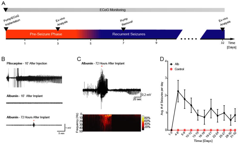
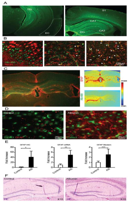
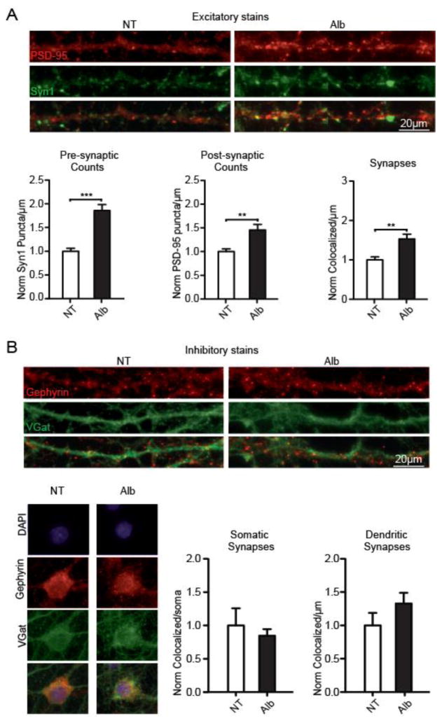
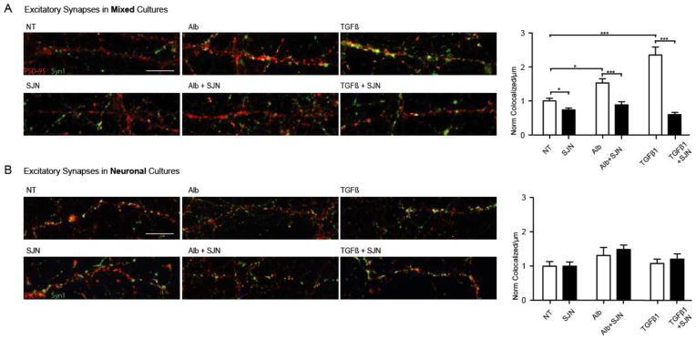
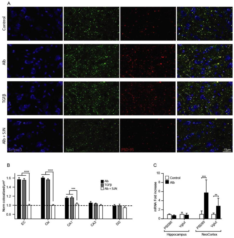
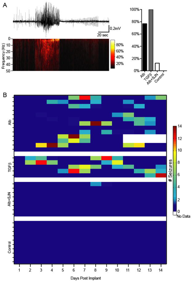
Post-injury epilepsy (PIE) is a common complication following brain insults, including ischemic, and traumatic brain injuries. At present, there are no means to identify the patients at risk to develop PIE or to prevent its development. Seizures can occur months or years after the insult, do not respond to anti-seizure medications in over third of the patients, and are often associated with significant neuropsychiatric morbidities. We have previously established the critical role of blood-brain barrier dysfunction in PIE, demonstrating that exposure of brain tissue to extravasated serum albumin induces activation of inflammatory transforming growth factor beta (TGF-β) signaling in astrocytes and eventually seizures. However, the link between the acute astrocytic inflammatory responses and reorganization of neural networks that underlie recurrent spontaneous seizures remains unknown. Here we demonstrate in vitro and in vivo that activation of the astrocytic ALK5/TGF-β-pathway induces excitatory, but not inhibitory, synaptogenesis that precedes the appearance of seizures. Moreover, we show that treatment with SJN2511, a specific ALK5/TGF-β inhibitor, prevents synaptogenesis and epilepsy. Our findings point to astrocyte-mediated synaptogenesis as a key epileptogenic process and highlight the manipulation of the TGF-β-pathway as a potential strategy for the prevention of PIE.

Keywords: ALK5; Albumin; Astrocytes; Blood–brain barrier (BBB); Epilepsy; Post-insult epilepsy (PIE); Post-traumatic epilepsy (PTE); Seizures; Synaptogenesis; TGF-β.


Conflict of interest statement

Competing interests:

The authors declare no competing interests.

Figures

Figure 1
Figure 1. ICV albumin induces latent appearance of spontaneous recurrent seizures
(A) The experimental paradigm in vivo. (B) Continuous ECoG recordings show that ICV albumin does not induce acute seizures or status epilepticus (common in other models of acquired epilepsy, e.g pilocarpine), yet results in latent appearance of spontaneous seizures. (C) Representative seizure recorded 72 h after pump implantation. (D) Comparison of number of seizures per day (averaged over 3 day periods) shows higher incidence of seizures in the albumin-infused mice (black line, n=13), compared to controls (red line, n=10). Notably, seizures appeared 3 days after initiating albumin infusion, and lasted well after the extraction of the pumps on day 7. Error bars indicate SEM.
Figure 2
Figure 2. ICV Albumin induces activation of astrocytes
(A) 72 hour infusion of fluorescein-conjugated albumin (10%; Alb-488, green) into the lateral ventricle results in florescence accumulation in the hippocampus (DG, CA1,3), the somatosensory cortex (S1), corpus callosum, entorhinal cortex (EC) and adjacent neocortical regions, as evident here in coronal slices 72h after pump implantation (right,~ bregma −1.58mm; left,~bregma −2.92mm). (B) Best representative images of albumin (Alb-488) co-localization with neurons (NeuN, somatosensory cortex), microglia (IBA1, hippocampal hilus) and astrocytes (GFAP, hippocampal hilus). White arrowheads mark co-localization of cells with fluorescein-conjugated albumin. (C) GFAP immunohistochemical staining of a coronal slice, 72h after infusion of fluorescein-conjugated albumin. GFAP and albumin florescent intensity maps (right panels) show high levels of fluorescence in the hippocampus, the somatosensory cortex, corpus callosum and adjacent neocortical regions. (D) Immunohistochemical (IHC) staining of cell nuclei (ToPro3, green) and GFAP positive cells (red) demonstrates increased levels of GFAP following ICV albumin treatment in the hilus of the hippocampus. (E) Quantitative analyses of IHC staining (IHC, ctrl=4 alb=5) of the hippocampus reveals a significant increase in GFAP expression, further confirmed by real time RT-PCR (mRNA, ctrl=3 alb=3), and western blots (western, ctrl=4 alb=6). (F) Hematoxylin eosin (HE) hippocampal staining shows no gross abnormalities one month after albumin infusion, compared to controls (ctrl=3 alb=3). Error bars indicate SEM. *p ≤ 0.05, **p ≤ 0.01, ***p ≤ 0.001.
Figure 3
Figure 3. Albumin induces excitatory, but not inhibitory, synaptogenesis in temporal cultures
(A) Staining for excitatory presynaptic (Syn1; green) and postsynaptic (PSD-95, red) proteins, revealed a significant albumin-induced increase in presynaptic, postsynaptic and co-localized counts along dendritic lengths (Alb=17; no treatment (NT)=12). (B) Staining for inhibitory presynaptic (V-Gat, green) and postsynaptic (Gephyrin, red) proteins showed no significant albumin-induced differences in synaptic counts, quantified along dendritic lengths and the somatic area (per soma: Alb=17; NT=15; p=0.2569; and per dendrite Alb=19; NT=18. P=0.0854). Error bars indicate SEM. *p ≤ 0.05, **p ≤ 0.01, ***p ≤ 0.001.
Figure 4
Figure 4. Albumin-induced excitatory synaptogenesis is mediated by astrocyte-driven TGF-β\ALK5 signaling
(A) In mixed cultures of astrocytes and neurons, 72 hour TGF-β1 incubation (n=18) induced a synaptogenic effect similar to that of albumin (n=17), with both treatments resulting in significantly higher synaptic counts compared to untreated controls (no treatment (NT)=17). Both albumin- and TGF-β1- induced synaptogenesis was significantly blocked by the selective TGF-β type I receptor ALK5 inhibitor (SJN2511, SJN) (NT=15;Alb+SJN=21;TGFβ+SJN=20). (B) No significant increase in synaptic counts was observed in enriched neuronal cultures (P>0.6; Alb=13; TGFβ=14; NT=15), and pre-incubation with SJN had no effect on synaptic counts in the absence of astrocytes (P>0.9; SJN=15; SJN+Alb=13; SJN+TGFβ1=13). Error bars indicate SEM. *p ≤ 0.05, **p ≤ 0.01, ***p ≤ 0.001.
Figure 5
Figure 5. Albumin exposure in-vivo is associated with region-specific, TGF-β1 mediated synaptogenesis
(A) Representative z-stacked images of pre-synaptic (Syn1, green) and post-synaptic (PSD-95) markers 72 h after pump implantation. (B) Synaptic quantification demonstrates that TGF-β1 (n=3 × 6 slices) and albumin (n=4 × 6 slices) induce similar synaptogenic effects, blocked by SJN2511. (C) RT-PCR demonstrates a significant increase in cortical expression of post-synaptic (PSD95) and pre-synaptic (Vglut) protein mRNA in albumin treated mice (n=8), compared to controls (n=12). Error bars indicate SEM. *p ≤ 0.05, **p ≤ 0.01, ***p ≤ 0.001.
Figure 6
Figure 6. Albumin-induced seizures are mediated by TGF-β and can be prevented with SJN2511
(A) ICV TGF-β induces spontaneous epileptic seizures, comparable to those observed following ICV albumin. However, while albumin induced epilepsy in 77% of mice (n=13), all TGF-β-infused mice developed seizures (n=5). Moreover, co-infusion with SJN2511 (n=8) prevented seizures in all but one animal. (B) Albumin and TGF-β-induced seizures persisted throughout the monitoring period (and well after pump extraction on day 7). The one epileptic animal in the SJN group suffered two seizures on day 9.

Similar articles

Cited by

References

    1. Abbott NJ, Friedman A. Overview and introduction: the blood-brain barrier in health and disease. Epilepsia. 2012;53(Suppl 6):1–6. doi: 10.1111/j.1528-1167.2012.03696.x. - DOI - PMC - PubMed
    1. Allen NJ, Bennett ML, Foo LC, Wang GX, Chakraborty C, Smith SJ, Barres BA. Astrocyte glypicans 4 and 6 promote formation of excitatory synapses via GluA1 AMPA receptors. Nature. 2012;486:410–4. - PMC - PubMed
    1. Aronica E, Boer K, Becker A, Redeker S, Spliet WG, van Rijen PC, Wittink F, Breit T, Wadman WJ, Lopes da Silva FH, Troost D, Gorter JA. Gene expression profile analysis of epilepsy-associated gangliogliomas. Neuroscience. 2008;151:272–292. doi: 10.1016/j.neuroscience.2007.10.036. - DOI - PubMed
    1. Babb TL, Kupfer WR, Pretorius JK, Crandall PH, Levesque MF. Synaptic reorganization by mossy fibers in human epileptic fascia dentata. Neuroscience. 1991;42:351–363. - PubMed
    1. Balosso S, Maroso M, Sanchez-Alavez M, Ravizza T, Frasca A, Bartfai T, Vezzani A. A novel non-transcriptional pathway mediates the proconvulsive effects of interleukin-1beta. Brain. 2008;131:3256–65. doi: 10.1093/brain/awn271. - DOI - PMC - PubMed

Publication types

MeSH terms

Substances