. 2015 Jun:30:11-20.
doi: 10.1016/j.dnarep.2015.03.002. Epub 2015 Mar 17.

BRCA1 and BRCA2 protect against oxidative DNA damage converted into double-strand breaks during DNA replication

Affiliations

BRCA1 and BRCA2 protect against oxidative DNA damage converted into double-strand breaks during DNA replication

Ram Fridlich et al. DNA Repair (Amst). 2015 Jun.

Abstract

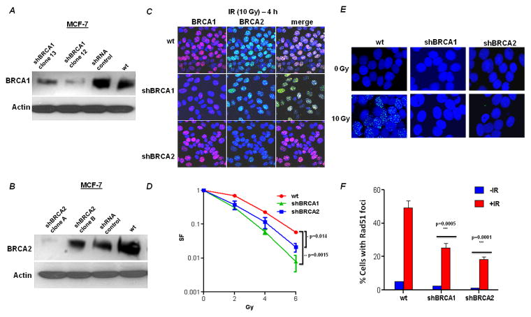
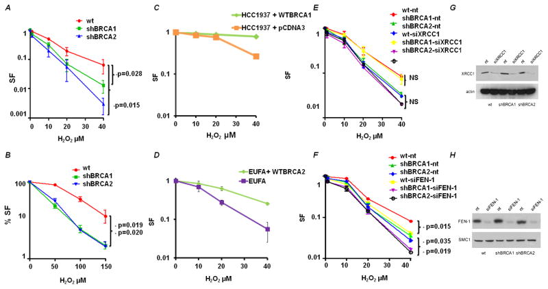
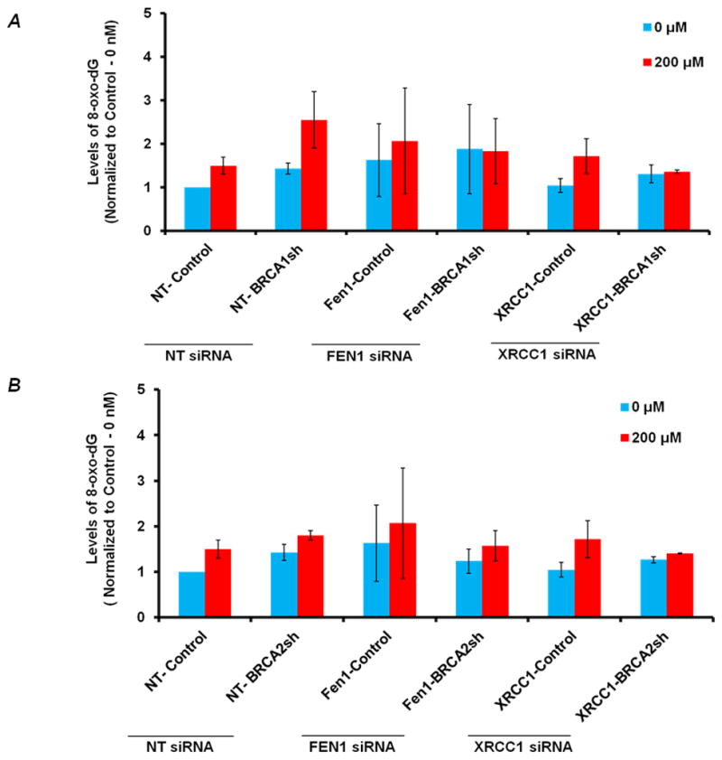
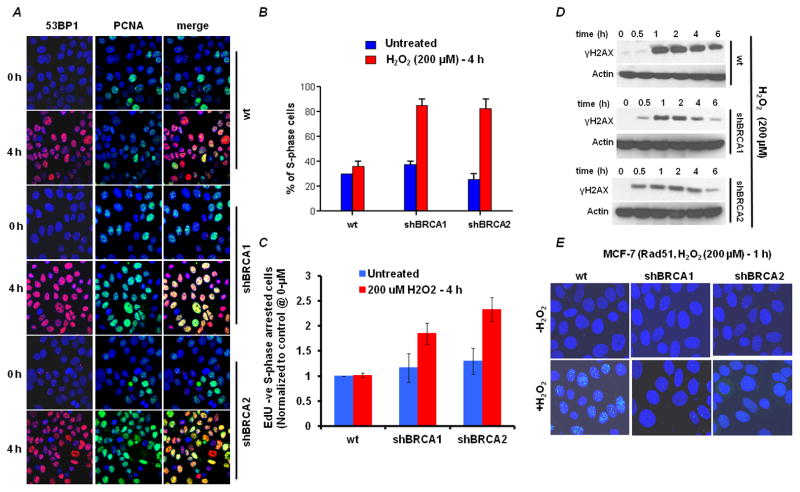
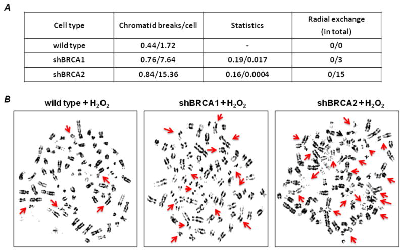
BRCA1 and BRCA2 mutation carriers are predisposed to develop breast and ovarian cancers, but the reasons for this tissue specificity are unknown. Breast epithelial cells are known to contain elevated levels of oxidative DNA damage, triggered by hormonally driven growth and its effect on cell metabolism. BRCA1- or BRCA2-deficient cells were found to be more sensitive to oxidative stress, modeled by treatment with patho-physiologic concentrations of hydrogen peroxide. Hydrogen peroxide exposure leads to oxidative DNA damage induced DNA double strand breaks (DSB) in BRCA-deficient cells causing them to accumulate in S-phase. In addition, after hydrogen peroxide treatment, BRCA deficient cells showed impaired Rad51 foci which are dependent on an intact BRCA1-BRCA2 pathway. These DSB resulted in an increase in chromatid-type aberrations, which are characteristic for BRCA1 and BRCA2-deficient cells. The most common result of oxidative DNA damage induced processing of S-phase DSB is an interstitial chromatid deletion, but insertions and exchanges were also seen in BRCA deficient cells. Thus, BRCA1 and BRCA2 are essential for the repair of oxidative DNA damage repair intermediates that persist into S-phase and produce DSB. The implication is that oxidative stress plays a role in the etiology of hereditary breast cancer.

Keywords: BRCA; Cancer; Chromosome aberrations; Homologous recombination; Oxidative stress.


Conflict of interest statement

Conflict of Interest Statement

Simon Powell have no financial conflicts of interest with any of the work in this submitted paper.

Simon Powell have a single position of employment (Memorial Sloan-Kettering) and no other sources of income.

Figures

Figure 1
Figure 1. Depletion of BRCA1 and BRCA2 by shRNA in MCF-7 cells
MCF-7 cells were stably transfected with empty vector (pKLO.1) or with short hairpin RNA constructs against BRCA1 or BRCA2. The efficacy of the shRNA constructs in depleting BRCA1 (A) and BRCA2 (B) was determined by immunoblotting (actin served as loading control) and confirmed by immunofluorescence. (C) Confocal microscopic images of cell clones using immunofluorescent staining for BRCA1 and BRCA2. Wild type (empty vector) and BRCA1 or BRCA2 deficient MCF-7 cells were irradiated (10 Gy) and 4h later, the cells were fixed and processed for BRCA1 (red) or BRCA2 (green) immunofluorescence. The nuclei were identified by DAPI staining (blue). (D) Relative survival of BRCA1- or BRCA2-deficient cells after exposure to IR. Data are the mean and SE of the mean from three independent experiments. The survival of BRCA1- and BRCA2- deficient cells relative to wild type MCF-7 at 6 Gy was compared for statistical significance. (E) Formation of Rad51 foci (green) in response to DNA damage induced by IR. Nuclei were stained with DAPI. Representative nuclei are displayed from either untreated or irradiated cells. Quantification of the percentage of foci positive cells is shown (mean and SE of the mean from three independent experiments).
Figure 2
Figure 2. BRCA1- and BRCA2-deficient cells are sensitive to H2O2
(A) Clonogenic survival of BRCA-deficient cells after exposure to H2O2. Data shown are the mean and SE of the mean from three independent experiments. The survival of BRCA1 and BRCA2 deficient cells relative to wild type MCF-7 at 40μM was compared for statistical significance. (B) MTS assay of BRCA-deficient cells after exposure to H2O2. (C) Clonogenic survival of BRCA1-deficient cell line – HCC1937 containing pcDNA3 empty vector and HCC137 complemented with a full-length wtBRCA1 construct after exposure to H2O2. Data shown are the mean and SE from three independent experiments. (D) Clonogenic survival of BRCA2-deficient cell line – EUFA and EUFA complemented with a full-length wtBRCA2 construct after exposure to H2O2. Data shown are the mean and SE from three independent experiments. (E) XRCC1-depletion in wild type and BRCA-deficient cells. Cells were harvested 48h after siXRCC1 or control siRNA (nt) nucleofection and cell lysates were immuno-blotted for XRCC1. Clonogenic survival of wild type and BRCA deficient cells nucleotransfected with siXRCC1 or control siRNA (nt) after exposure to H2O2 as in A. (F) The effect of FEN1-depletion as in E. (G) Representative immunoblots confirming XRCC1 depletion in BRCA1 and BRCA2 deficient cells. (H) Representative immunoblots confirming FEN1 depletion in BRCA1 and BRCA2 deficient cells. Actin was used as loading control. Cell lines were nucleotransfected with FEN1/XRRC1 siRNA and post 48 hrs from transfection, cells were harvested for immunoblotting.
Figure 3
Figure 3. A: Levels of 8-oxo-dG in BRCA1-depleted and FEN1-BRCA1 or XRCC1-BRCA1 co-depleted cells.<
br>The relative levels of 8-oxo-dG (Mean Fluorescence Intensity, MFI –normalized to 0-nM of control cells) in BRCA1-depleted, FEN1-BRCA1 and XRCC1-BRCA1 co-depleted cells respectively before and after H2O2 treatment. Cells were nucleofected with FEN1 and XRCC1 siRNA respectively and 48 hrs after transfection treated with 200μM of H2O2 for 1 hr. Subsequently, harvested cells were subjected to 8-oxo-dG analysis by flow cytometry. 8-oxodG was determined by using primary antibody against 8-oxodG (Trevigen) and Alexa fluor 488-Rabbit-anti-Mouse secondary antibody IgG (H+L) (Molecular Probes®) using FACS (LSRII). Results are from three independent experiments and are expressed as the mean ± SE. To reduce the basal levels of oxidative DNA damage cells were incubated in 3% Oxygen incubator. B: Levels of 8-oxo-dG in BRCA2-depleted and FEN1-BRCA2 or XRCC1-BRCA2 co-depleted cells: Relative levels of 8-oxo-dG (Mean Fluorescence Intensity, MFI – normalized to 0-nM of control cells) in BRCA2-depleted, FEN1-BRCA2 and XRCC1-BRCA2 co-depleted cells respectively before and after H2O2 treatment as in A.
Figure 4
Figure 4. BRCA-deficient cells accumulate in S phase due to oxidative stress-induced double-strand breaks
(A) Cells were treated with 200μM H2O2 for 4h and the immunofluorescence staining pattern was imaged. Double-strand breaks are shown by 53BP1 (red); S-phase cells are shown by the presence of PCNA in nuclear foci (green). (B) Quantification of the percentage of PCNA foci-positive cells from A. Data shown are the mean and SE from three independent experiments. (C) Quantification of S phase arrested BRCA deficient cells. Cells were treated with 200μM H2O2 for 4h and flow-cytometric analysis of S phase arrested cells after exposure to H2O2 treatment (EdU negative – propidium iodide S phase DNA content) showing reduced incorporation of EdU were measured (additional data in Fig S3). Flow cytometric data is normalized to untreated control cells. Data shown are the mean and SE from three independent experiments. (D) Representative Immunoblots of γH2AX accumulation in BRCA1, BRCA2 deficient and control cells. Actin was used as loading control. Cell lines were treated with 200μM H2O2 for shown time points and were harvested for immunoblotting. (E) Formation of Rad51 foci (green) 4h after treatment with 200μM H2O2 for 1h.
Figure 5
Figure 5. H2O2 induces chromosome aberrations in BRCA1- and BRCA2-deficient cells
Untreated and H2O2-treated cells were subjected to cytogenetic analysis. (A) Chromatid breaks are shown as the average number of breaks per cell. Spontaneous aberrations were derived from the analysis of 50 metaphase spreads and are shown as the first result for each cell type; the second result is following treatment with H2O2. Statistical comparisons are shown for BRCA1- and BRCA2-deficient cells compared with wild type cells. Radial exchanges are shown in the fourth column. (B) Representative images of H2O2-treated metaphase spreads. Arrows indicate chromatid/chromosome breaks and radial exchanges in each cell type. (See additional details in Supplementary Figure 2).

Similar articles

Cited by

References

    1. McCord JM. Superoxide dismutase in aging and disease: an overview. Methods Enzymol. 2002;349:331–41. - PubMed
    1. Brown NS, Bicknell R. Hypoxia and oxidative stress in breast cancer. Oxidative stress: its effects on the growth, metastatic potential and response to therapy of breast cancer. Breast Cancer Res. 2001;3(5):323–7. - PMC - PubMed
    1. Hursting SD, et al. Mechanism-based cancer prevention approaches: targets, examples, and the use of transgenic mice. J Natl Cancer Inst. 1999;91(3):215–25. - PubMed
    1. Lavigne JA, et al. The effects of catechol-O-methyltransferase inhibition on estrogen metabolite and oxidative DNA damage levels in estradiol-treated MCF-7 cells. Cancer Res. 2001;61(20):7488–94. - PubMed
    1. Hamada J, et al. Increased oxidative DNA damage in mammary tumor cells by continuous epidermal growth factor stimulation. J Natl Cancer Inst. 2001;93(3):214–9. - PubMed

Publication types