. 2015 Apr 5;128(7):919-27.
doi: 10.4103/0366-6999.154298.

Propofol ameliorates calpain-induced collapsin response mediator protein-2 proteolysis in traumatic brain injury in rats

Affiliations

Propofol ameliorates calpain-induced collapsin response mediator protein-2 proteolysis in traumatic brain injury in rats

Yun Yu et al. Chin Med J (Engl). .

Abstract

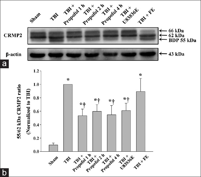
Background: Collapsin response mediator protein-2 (CRMP2), a multifunctional cytosolic protein highly expressed in the brain, is degraded by calpain following traumatic brain injury (TBI), possibly inhibiting posttraumatic neurite regeneration. Lipid peroxidation (LP) is involved in triggering postinjury CRMP2 proteolysis. We examined the hypothesis that propofol could attenuate LP, calpain-induced CRMP2 degradation, and brain injury after TBI.

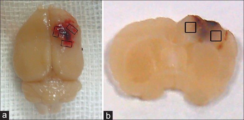
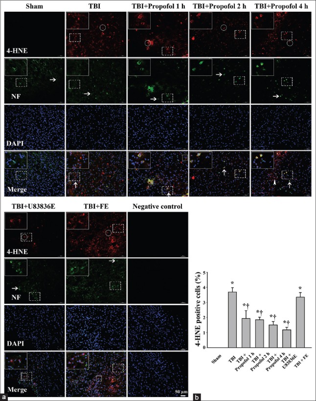
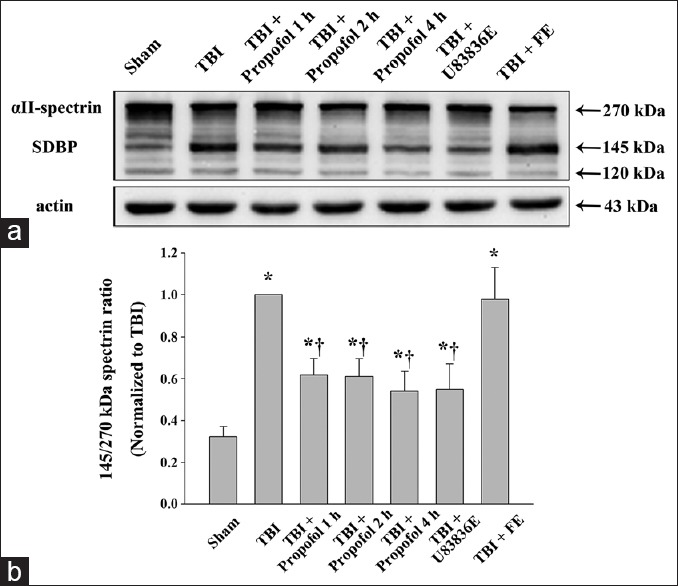
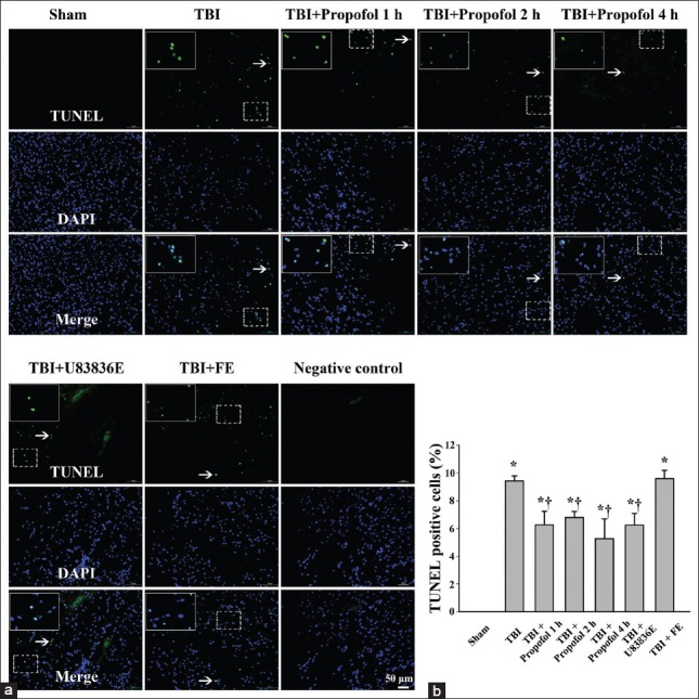
Methods: A unilateral moderate controlled cortical impact injury was induced in adult male Sprague-Dawley rats. The animals were randomly divided into seven groups: Sham control group, TBI group, TBI + propofol groups (including propofol 1 h, 2 h, and 4 h groups), TBI + U83836E group and TBI + fat emulsion group. The LP inhibitor U83836E was used as a control to identify that antioxidation partially accounts for the potential neuroprotective effects of propofol. The solvent of propofol, fat emulsion, was used as the vehicle control. Ipsilateral cortex tissues were harvested at 24 h post-TBI. Immunofluorescent staining, Western blot analysis, and terminal deoxynucleotidyl transferase-mediated dUTP nick-end labeling were used to evaluate LP, calpain activity, CRMP2 proteolysis and programmed cell death. The data were statistically analyzed using one-way analysis of variance and a paired t-test.

Results: Propofol and U83836E significantly ameliorated the CRMP2 proteolysis. In addition, both propofol and U83836E significantly decreased the ratio of 145-kDa αII-spectrin breakdown products to intact 270-kDa spectrin, the 4-hydroxynonenal expression and programmed cell death in the pericontusional cortex at 24 h after TBI. There was no difference between the TBI group and the fat emulsion group.

Conclusions: These results demonstrate that propofol postconditioning alleviates calpain-mediated CRMP2 proteolysis and provides neuroprotective effects following moderate TBI potentially by counteracting LP and reducing calpain activation.


Conflict of interest statement

Conflict of Interest: None declared.

Figures

Figure 1
Figure 1
Schematic representation of the sample area for detection. (a) Image of a representative TBI brain showing cortex contusion and the pericontusional region surrounding the injured cortex. The sample area for detection is marked with the black boxes. TBI: Traumatic brain injury; (b) a coronal section of a rat brain used for immunofluorescent staining and TUNEL analysis. The black boxes indicate the detected region. TUNEL: Terminal deoxynucleotidyl transferase-mediated dUTP nick-end labeling.
Figure 2
Figure 2
Immunofluorescence staining of 4-hydroxynonenal positive cells in the pericontusional cortex at 24 h following moderate TBI in the Sham group (Sham), TBI group (TBI), three groups treated with propofol at different time points after TBI (TBI + Propofol 1 h, TBI + Propofol 2 h, TBI + Propofol 4 h), the lipid peroxidation inhibitor U83836E group (TBI + U83836E) and the vehicle control fat emulsion group (TBI + FE). TBI: Traumatic brain injury; (a) Horizontal arrows indicate representative neurofilaments (NF; green). Round rings and arrowheads indicate representative 4-HNE-modified proteins (red) expressed in the cytoplasm and the nucleus, respectively. Vertical arrows and boxes represent 4-HNE-positive neurons and non-neuronal cells, respectively; (b) quantitation of 4-HNE-positive cells. The data are presented as the mean ± standard deviation (SD) (n = 3). *P < 0.01 vs. Sham group; P < 0.01 vs. TBI group. Bar = 50 μm.
Figure 3
Figure 3
Western blot analysis of αII-spectrin in the pericontusional cortex at 24 h following moderate TBI. (a) Brain tissue lysates were immunoblotted with anti-αII-spectrin (top) and anti-β-actin (bottom) antibodies, respectively. The lanes were loaded with protein from the Sham group (Sham), the TBI group (TBI), the propofol 1 h group (TBI + Propofol 1 h), the propofol 2 h group (TBI + Propofol 2 h), the propofol 4 h group (TBI + Propofol 4 h), the lipid peroxidation inhibitor U83836E group (TBI + U83836E) and the vehicle control fat emulsion group (TBI + FE). TBI: Traumatic brain injury; (b) quantitative analysis for the ratio of the calpain-mediated 145-kDa spectrin breakdown product (SBDP) to intact 270-kDa αII-spectrin. The densitometric ratio was normalized against the TBI group. The results were expressed as the means ± standard deviation (SD) (n = 6). *P < 0.01 vs. Sham group; P < 0.01 vs. TBI group.
Figure 4
Figure 4
Western blot analysis of collapsin response mediator protein-2 (CRMP2) proteolysis in the ipsilateral cortex surrounding the impacted area at 24 h following moderate TBI. (a) Brain tissue lysates were immunoblotted with anti-CRMP2 (top) and anti-β-actin (bottom) antibodies. The lanes were loaded with protein from the Sham group (Sham), the TBI group (TBI), the propofol 1 h group (TBI + Propofol 1 h), the propofol 2 h group (TBI + Propofol 2 h), the propofol 4 h group (TBI + Propofol 4 h), the lipid peroxidation inhibitor U83836E group (TBI + U83836E) and the vehicle control fat emulsion group (TBI + FE). TBI: Traumatic brain injury; (b) densitometric analysis for the ratio of the 55-kDa breakdown product to intact 62-kDa CRMP2. The densitometric ratio was normalized against the TBI group. The data are expressed as the means ± standard deviation (SD) (n = 6). *P < 0.01 vs. Sham group; †P < 0.01 vs. TBI group.
Figure 5
Figure 5
Terminal deoxynucleotidyl transferase-mediated dUTP nick-end labeling (TUNEL) of programmed cell death in the pericontusional cortex at 24 h following moderate TBI in the Sham group (Sham), TBI group (TBI), three propofol groups (TBI + Propofol 1 h, TBI + Propofol 2 h, TBI + Propofol 4 h), lipid peroxidation (LP) inhibitor U83836E group (TBI + U83836E) and the vehicle control fat emulsion group (TBI + FE). TUNEL labeling demonstrated the reduction in programmed cell death after the administration of propofol and U83836E following TBI. TBI: Traumatic brain injury; TUNEL: Terminal deoxynucleotidyl transferase-mediated dUTP nick-end labeling. (a) The white arrows indicate the TUNEL-positive cells, which represent programmed cell death; (b) quantitation of TUNEL-positive cells. The results are presented as the means ± standard deviation (SD) (n = 3). *P < 0.01 vs. Sham group; P < 0.01 vs. TBI group. Bar = 50 μm.

Similar articles

Cited by

References

    1. Georgiou AP, Manara AR. Role of therapeutic hypothermia in improving outcome after traumatic brain injury: A systematic review. Br J Anaesth. 2013;110:357–67. - PubMed
    1. Mustafa AG, Singh IN, Wang J, Carrico KM, Hall ED. Mitochondrial protection after traumatic brain injury by scavenging lipid peroxyl radicals. J Neurochem. 2010;114:271–80. - PMC - PubMed
    1. Luo T, Wu J, Kabadi SV, Sabirzhanov B, Guanciale K, Hanscom M, et al. Propofol limits microglial activation after experimental brain trauma through inhibition of nicotinamide adenine dinucleotide phosphate oxidase. Anesthesiology. 2013;119:1370–88. - PubMed
    1. Boer C, Franschman G, Loer SA. Prehospital management of severe traumatic brain injury: Concepts and ongoing controversies. Curr Opin Anaesthesiol. 2012;25:556–62. - PubMed
    1. Hall ED, Vaishnav RA, Mustafa AG. Antioxidant therapies for traumatic brain injury. Neurotherapeutics. 2010;7:51–61. - PMC - PubMed

Publication types