

In vivo confocal microscopic observation of lamellar corneal transplantation in the rabbit using xenogenic acellular corneal scaffolds as a substitute
- PMID: 25836615
- PMCID: PMC4834011
- DOI: 10.4103/0366-6999.154301
In vivo confocal microscopic observation of lamellar corneal transplantation in the rabbit using xenogenic acellular corneal scaffolds as a substitute
Abstract
Background: The limiting factor to corneal transplantation is the availability of donors. Research has suggested that xenogenic acellular corneal scaffolds (XACS) may be a possible alternative to transplantation. This study aimed to investigate the viability of performing lamellar corneal transplantation (LCT) in rabbits using canine XACS.
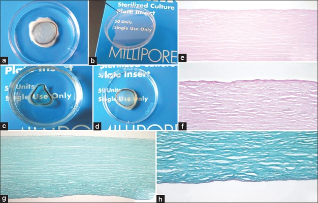
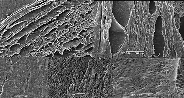
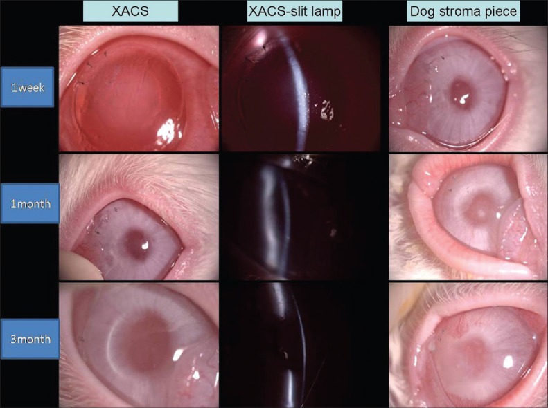
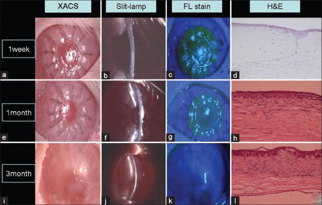
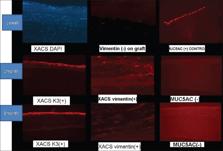
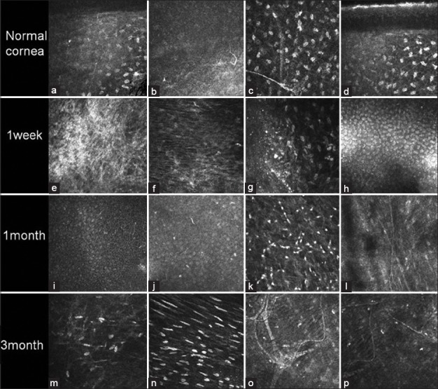
Methods: Fresh dog corneas were decellularized by serial digestion, and LCT was performed on rabbit eyes using xenogeneic decellularized corneal matrix. Cellular and morphological changes were observed by slit-lamp, light, and scanning electron microscopy at 7, 30 and 90 days postoperatively. Immunocytochemical staining for specific markers such as keratin 3, vimentin and MUC5AC, was used to identify cells in the graft.
Results: Decellularized xenogenic corneal matrix remained transparent for about 1-month after LCT. The recipient cells were able to survive and proliferate into the grafts. Three months after transplantation, grafts had merged with host tissue, and graft epithelialization and vascularization had occurred. Corneal nerve fibers were able to grow into the graft in rabbits transplanted with XACS.
Conclusions: Xenogenic acellular corneal scaffolds can maintain the transparency of corneal grafts about 1-month and permit growth of cells and nerve fibers, and is, therefore, a potential substitute or carrier for a replacement cornea.
Conflict of interest statement
Figures
Similar articles
-
Acellular human corneal matrix sheets seeded with human adipose-derived mesenchymal stem cells integrate functionally in an experimental animal model.Exp Eye Res. 2015 Mar;132:91-100. doi: 10.1016/j.exer.2015.01.020. Epub 2015 Jan 24. Exp Eye Res. 2015. PMID: 25625506
-
[Biocompatibility of acellular corneal stroma and transplantation of tissue-engineered corneal epithelium].Zhonghua Yan Ke Za Zhi. 2008 Oct;44(10):934-42. Zhonghua Yan Ke Za Zhi. 2008. PMID: 19176124 Chinese.
-
Biocompatibility and functionality of a tissue-engineered living corneal stroma transplanted in the feline eye.Invest Ophthalmol Vis Sci. 2014 Oct 2;55(10):6908-20. doi: 10.1167/iovs.14-14720. Invest Ophthalmol Vis Sci. 2014. PMID: 25277228
-
[Transplantation of corneal endothelial cells].Nippon Ganka Gakkai Zasshi. 2002 Dec;106(12):805-35; discussion 836. Nippon Ganka Gakkai Zasshi. 2002. PMID: 12610838 Review. Japanese.
-
Integration and remodelling of a collagen anterior lamellar keratoplasty graft in an animal model - A preliminary report.Exp Eye Res. 2021 Aug;209:108661. doi: 10.1016/j.exer.2021.108661. Epub 2021 Jun 5. Exp Eye Res. 2021. PMID: 34102207 Review.
Cited by
-
Chemical, Physical, and Biological Corneal Decellularization Methods: A Review of Literature.J Ophthalmol. 2024 Mar 25;2024:1191462. doi: 10.1155/2024/1191462. eCollection 2024. J Ophthalmol. 2024. PMID: 38567029 Free PMC article. Review.
-
Small Incision Femtosecond Laser-assisted X-ray-irradiated Corneal Intrastromal Xenotransplantation in Rhesus Monkeys: A Preliminary Study.Curr Mol Med. 2018;18(9):612-621. doi: 10.2174/1566524019666190129123935. Curr Mol Med. 2018. PMID: 30698112 Free PMC article.
-
Preliminary studies of constructing a tissue-engineered lamellar corneal graft by culturing mesenchymal stem cells onto decellularized corneal matrix.Int J Ophthalmol. 2021 Jan 18;14(1):10-18. doi: 10.18240/ijo.2021.01.02. eCollection 2021. Int J Ophthalmol. 2021. PMID: 33469478 Free PMC article.
-
A novel method in preparation of acellularporcine corneal stroma tissue for lamellar keratoplasty.Am J Transl Res. 2015 Dec 15;7(12):2612-29. eCollection 2015. Am J Transl Res. 2015. PMID: 26885261 Free PMC article.
-
Comparison of femtosecond laser-assisted corneal intrastromal xenotransplantation and the allotransplantation in rhesus monkeys.BMC Ophthalmol. 2017 Nov 9;17(1):202. doi: 10.1186/s12886-017-0595-z. BMC Ophthalmol. 2017. PMID: 29121878 Free PMC article.
References
-
- Auchincloss H, Jr, Sachs DH. Xenogeneic transplantation. Annu Rev Immunol. 1998;16:433–70. - PubMed
-
- Steele DJ, Auchincloss H., Jr Xenotransplantation. Annu Rev Med. 1995;46:345–60. - PubMed
-
- Auchincloss H., Jr Cell-mediated xenoresponses: Strong or weak? Clin Transplant. 1994;8:155–9. - PubMed
-
- Chapekar MS. Tissue engineering: Challenges and opportunities. J Biomed Mater Res. 2000;53:617–20. - PubMed
-
- Sano Y, Ksander BR, Streilein JW. Murine orthotopic corneal transplantation in high-risk eyes. Rejection is dictated primarily by weak rather than strong alloantigens. Invest Ophthalmol Vis Sci. 1997;38:1130–8. - PubMed
Publication types
MeSH terms
LinkOut - more resources
Full Text Sources
Other Literature Sources
