. 2015 Jun;156(6):2029-38.
doi: 10.1210/en.2014-1987. Epub 2015 Apr 2.

Reprogramming Mouse Cells With a Pancreatic Duct Phenotype to Insulin-Producing β-Like Cells

Affiliations

Reprogramming Mouse Cells With a Pancreatic Duct Phenotype to Insulin-Producing β-Like Cells

Takatsugu Yamada et al. Endocrinology. 2015 Jun.

Abstract

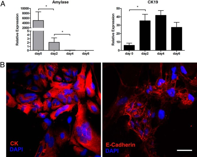
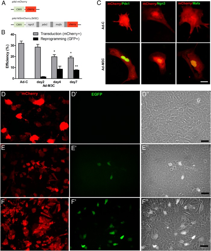
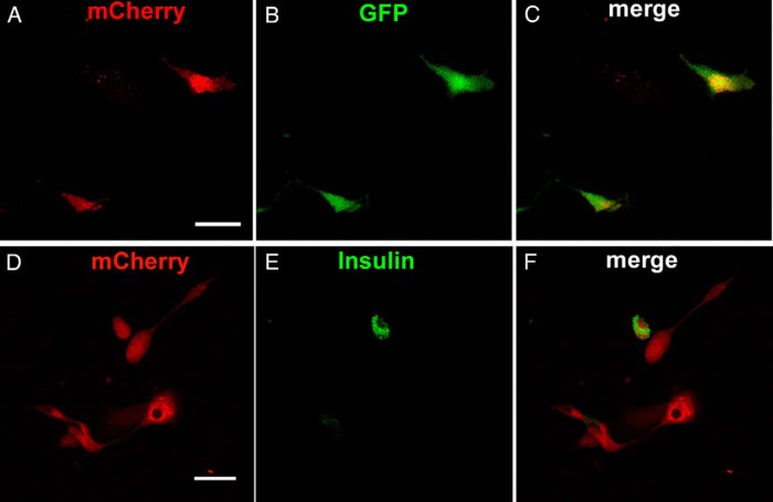
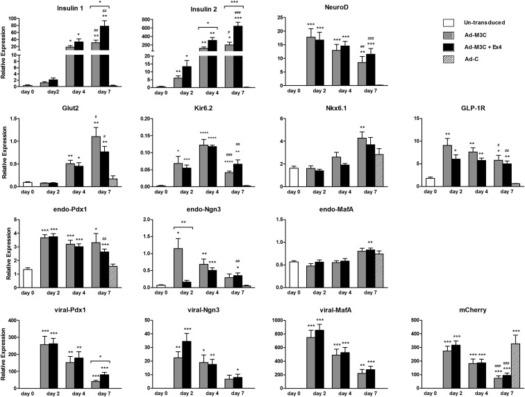
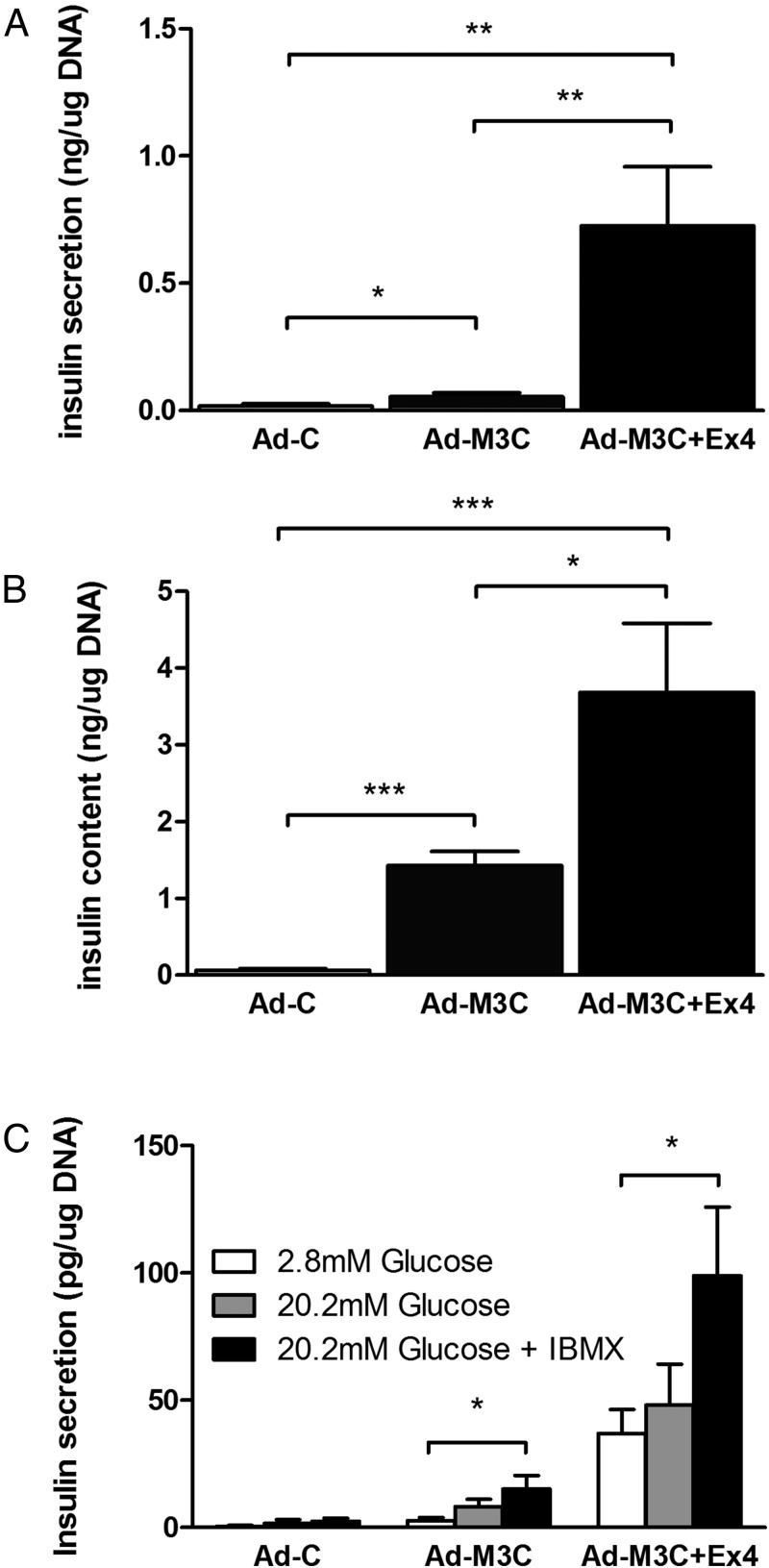
Reprogramming technology has opened the possibility of converting one cell type into another by forced expression of transgenes. Transduction of adenoviral vectors encoding 3 pancreatic transcription factors, Pdx1, Ngn3, and MafA, into mouse pancreas results in direct reprogramming of exocrine cells to insulin-producing β-like cells. We hypothesized that cultured adult pancreatic duct cells could be reprogrammed to become insulin-producing β-cells by adenoviral-mediated expression of this same combination of factors. Exocrine were isolated from adult mouse insulin 1 promoter (MIP)-green fluorescent protein (GFP) transgenic mice to allow new insulin-expressing cells to be detected by GFP fluorescence. Cultured cells were transduced by an adenoviral vector carrying a polycistronic construct Ngn3/Pdx1/MafA/mCherry (Ad-M3C) or mCherry sequence alone as a control vector. In addition, the effects of glucagon-like peptide-1 (GLP-1) receptor agonist, exendin-4 (Ex-4) on the reprogramming process were examined. GFP(+) cells appeared 2 days after Ad-M3C transduction; the reprogramming efficiency was 8.6 ± 2.6% by day 4 after transduction. Ad-M3C also resulted in increased expression of β-cell markers insulin 1 and 2, with enhancement by Ex-4. Expression of other β-cell markers, neuroD and GLP-1 receptor, were also significantly up-regulated. The amount of insulin release into the media and insulin content of the cells were significantly higher in the Ad-M3C-transduced cells; this too was enhanced by Ex-4. The transduced cells did not secrete insulin in response to increased glucose, indicating incomplete differentiation to β-cells. Thus, cultured murine adult pancreatic cells with a duct phenotype can be directly reprogrammed to insulin-producing β-like cells by adenoviral delivery of 3 pancreatic transcription factors.


Figures

Figure 1.
Figure 1.
Characterization of isolated exocrine cells. A, Changes in the gene expression profile of exocrine cells 0, 2, 4, and 6 days after isolation. Freshly isolated exocrine cells (d 0) had high expression of amylase, which disappeared in just 4 days. The results were obtained from the adherent cells after floating cells were removed on each day except day 0 (freshly isolated nonadherent exocrine cells). Mean ± SEM, 4 independent experiments (each with duplicates). *, P < .05. B, Seven days after isolation, the adherent cells had proliferated and formed epithelial-like monolayers with cobblestone-like morphology; immunostaining was for pan-CK (red) (left panel) and E-cadherin (red) (right panel). Blue represents nuclear staining with 4′,6-diamidino-2-phenylindole (DAPI). Scale bar, 50 μm. Images are representative of 4 independent experiments.
Figure 2.
Figure 2.
Adenoviral transduction of 3 transcriptional factors induced reprogramming of cells with duct phenotype to GFP+ β-like cells. A, Schematic constructs. CMV, cytomegaloviral promoter. Dark gray bar, 2A peptide, which mediates polycistronic protein expression. Cherry, monomeric cherry fluorescent protein. The construct pAd-M3m Cherry (M3C) consists of 4 different coding regions separated by 3 2A protein elements. Ad-C was prepared as the control. B, Transduction and reprogramming efficiency was assessed by quantification of the percentage of mCherry+ cells and GFP+ cells of total cells, respectively, using exocrine cells from MIP-GFP mice. Reprogrammed cells would be insulin-expressing GFP+ cells. Mean ± SEM, 3 independent experiments. *, P < .05; **, P < .001 compared with day 2. C, Expression of 3 transcription factors in Ad-C- and Ad-M3C-transduced cells 7 days after transduction of cells from DBA/2 mice. Transduced cells were marked by mCherry+ (red). Nuclear immunostaining of Pdx1, Ngn3, and MafA (green) was confirmed in cells transduced with Ad-M3C (bottom panels) but not detected in cells transduced with Ad-C (top panels). Images are representative of 3 independent experiments. Scale bar, 25 μm. D–F, The presence of mCherry and GFP was examined in the cells 2 days after transduction with Ad-C (D) or Ad-M3C (E) and then at 7 days after Ad-M3C (F) transduction. Transduced cells from MIP-GFP mice were marked by mCherry and some became GFP+ after Ad-M3C transduction. None of the mCherry+ cells in the Ad-C-transduced cells became GFP+. D”–F”, In all 3 conditions, the transduced GFP+ cells had a cobblestone-like morphology with differential interference contrast. Images are representative of 3 independent experiments. Scale bars, 25 μm (D) and 50 μm (E and F).
Figure 3.
Figure 3.
GFP and insulin expression in Ad-M3C-transduced cells 7 days after transduction. A–C, Coexpression of mCherry and GFP in cells from MIP-GFP mice fixed 7 days after Ad-M3C transduction and examined with confocal microscopy. Some mCherry+ (red)-transduced cells also expressed GFP fluorescence (green), indicating activity of the insulin promoter. Merged image (yellow). (D-F) Ad-M3C-treated mCherry+ (red) cells from DBA/2 mice were immunostained for insulin (green) 7 days after transduction. Merged images (yellow) showed coexpression of mCherry and insulin in some but not all Ad-M3C-transduced cells. Images are representative of 3 independent experiments. Scale bars, 50 μm.
Figure 4.
Figure 4.
Gene expression of phenotypic duct cells after Ad-M3C transduction. Changes in gene expression by Ad-M3C-transduction with and without Ex-4 were determined: untransduced cells as day 0, Ad-M3C-treated cells and Ad-M3C-treated plus Ex-4 (Ad-M3C+Ex4) cells on days 2, 4, and 7 of transduction, and Ad-C-treated cells on day 7. The 3 viral transgenes are referred to as viral-Pdx1, viral-Ngn3, and viral-MafA, whereas the 3 endogenous transcription factors are referred as endo-Pdx1, endo-Ngn3, and endo-MafA for encoding untranslated regions (UTRs). Mean ± SEM, 12 independent experiments (each with duplicates). *, P < .05; **, P < .01; ***, P < .001 compared with day 0; #, P < .05; ##, P < .01; ###, P < .001 compared with Ad-C on day 7. Brackets show the comparison between Ad-M3C and Ad-M3C+Ex4, *, P < .05; **, P < .01; ***, P < .001.
Figure 5.
Figure 5.
Ex-4 enhanced both insulin secretion and content of cells transduced with Ad-M3C. Ad-C-transduced cells, Ad-M3C-transduced cells, and Ad-M3C-transduced cells cultured with Ex-4 for 7 days, then the amount of insulin secreted into the media (A) and the insulin content of the cells (B) were measured by ELISA. Media were changed every 24 hours, and insulin levels were measured just before media change. Values were normalized to the total amount of DNA content in each well. Mean ± SEM, 8 independent experiments (each with duplicates); *, P < .05; **, P < .01; ***, P < .001 compared with Ad-C. C, GSIS in static incubation of Ad-C-treated PDCs, Ad-M3C-treated PDCs, and Ad-M3C-treated PDCs in combination with Ex-4 at day 7 of transduction. Cells were incubated for 1 hour either with low glucose (2.8mM), high glucose (20.2mM), or high glucose with 1-methyl-3-isobutylxanthine (IBMX) (1mM). Mean ± SEM, 4 independent experiments (each with duplicates). *, P < .05; **, P < .01.

References

    1. Weir GC, Bonner-Weir S. Islet β cell mass in diabetes and how it relates to function, birth, and death. Ann NY Acad Sci. 2013;1281:92–105. - PMC - PubMed
    1. Alejandro R, Barton FB, Hering BJ, Wease S. 2008 update from the Collaborative Islet Transplant Registry. Transplantation. 2008;86(12):1783–1788. - PubMed
    1. Kroon E, Martinson LA, Kadoya K, et al. Pancreatic endoderm derived from human embryonic stem cells generates glucose-responsive insulin-secreting cells in vivo. Nat Biotechnol. 2008;26(4):443–452. - PubMed
    1. Rezania A, Bruin JE, Riedel MJ, et al. Maturation of human embryonic stem cell-derived pancreatic progenitors into functional islets capable of treating pre-existing diabetes in mice. Diabetes. 2012;61(8):2016–2029. - PMC - PubMed
    1. Alipio Z, Liao W, Roemer EJ, et al. Reversal of hyperglycemia in diabetic mouse models using induced-pluripotent stem (iPS)-derived pancreatic β-like cells. Proc Natl Acad Sci USA. 2010;107(30):13426–13431. - PMC - PubMed

Publication types

Substances