

Visualization of the Peripheral Branches of the Mandibular Division of the Trigeminal Nerve on 3D Double-Echo Steady-State with Water Excitation Sequence
- PMID: 25836724
- PMCID: PMC7965273
- DOI: 10.3174/ajnr.A4288
Visualization of the Peripheral Branches of the Mandibular Division of the Trigeminal Nerve on 3D Double-Echo Steady-State with Water Excitation Sequence
Abstract
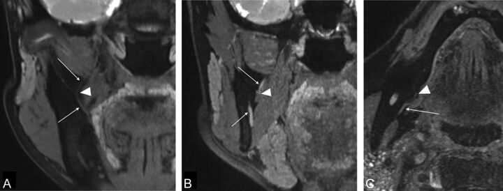
Background and purpose: Although visualization of the extracranial branches of the cranial nerves has improved with advances in MR imaging, only limited studies have assessed the detection of extracranial branches of the mandibular nerve (V3). We investigated the detectability of the branches of V3 on a 3D double-echo steady-state with water excitation sequence.
Materials and methods: We retrospectively evaluated the detectability of the 6 branches of the V3, the masseteric, buccal, auriculotemporal, lingual, inferior alveolar, and mylohyoid nerves, by using a 5-point scale (4, excellent; 3, good; 2, fair; 1, poor; and 0, none) in 86 consecutive patients who underwent MR imaging with the 3D double-echo steady-state with water excitation sequence. Weighted κ analysis was used to calculate interobserver variability among the 3 readers.
Results: The detection of the lingual and inferior alveolar nerves was the most successful, with excellent average scores of 3.80 and 3.99, respectively. The detection of the masseteric, the buccal, and the auriculotemporal nerves was good, with average scores of 3.31, 2.67, and 3.11, respectively. The mylohyoid nerve was difficult to detect with poor average scores of 0.62. All nerves had excellent interobserver variability across the 3 readers (average weighted κ value, 0.95-1.00).
Conclusions: The 3D double-echo steady-state with water excitation sequence demonstrated excellent visualization of the extracranial branches of V3 in most patients. The 3D double-echo steady-state with water excitation sequence has the potential for diagnosing V3 pathologies and preoperatively identifying peripheral cranial nerves to prevent surgical complications.
© 2015 by American Journal of Neuroradiology.
Figures

References
-
- Borges A, Casselman J. Imaging the cranial nerves. Part I. Methodology, infectious and inflammatory, traumatic and congenital lesions. Eur Radiol 2007;17:2112–25 - PubMed
-
- Borges A, Casselman J. Imaging the cranial nerves. Part II. Primary and secondary neoplastic conditions and neurovascular conflicts. Eur Radiol 2007;17:2332–44 - PubMed
MeSH terms
LinkOut - more resources
Full Text Sources
Medical