. 2015 Jul:76:58-66.
doi: 10.1016/j.bone.2015.03.019. Epub 2015 Mar 30.

In vivo mechanical loading rapidly activates β-catenin signaling in osteocytes through a prostaglandin mediated mechanism

Affiliations

In vivo mechanical loading rapidly activates β-catenin signaling in osteocytes through a prostaglandin mediated mechanism

N Lara-Castillo et al. Bone. 2015 Jul.

Abstract

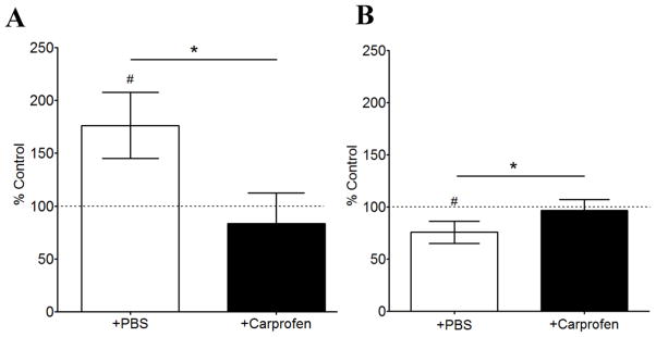
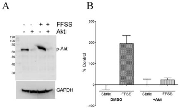
The response of the skeleton to loading appears to be mediated through the activation of the Wnt/β-catenin signaling pathway and osteocytes have long been postulated to be the primary mechanosensory cells in bone. To examine the kinetics of the mechanoresponse of bone and cell types involved in vivo, we performed forearm loading of 17-week-old female TOPGAL mice. β-catenin signaling was observed only in embedded osteocytes, not osteoblasts, at 1h post-loading, spreading to additional osteocytes and finally to cells on the bone surface by 24h. This early activation at 1h appeared to be independent of receptor (Lrp5/6) mediated activation as it occurred in the presence of the inhibitors sclerostin and/or Dkk1. The COX-2 inhibitor, Carprofen, blocked the activation of β-catenin signaling and decline in sclerostin positive osteocytes post-loading implying an important role for prostaglandin. In vitro, PI3K/Akt activation was shown to be required for β-catenin nuclear translocation downstream from prostaglandin in MLO-Y4 osteocyte-like cells supporting this mechanism. Downstream targets of β-catenin signaling, sclerostin and Dkk1, were also examined and found to be significantly downregulated in osteocytes in vivo at 24h post-loading. The pattern of initially activated osteocytes appeared random and in order to understand this heterogeneous expression, a novel finite element model of the strain field in the ulna was developed, which predicts highly variable local magnitudes of strain experienced by osteocytes. In summary, both in vivo and in vitro models show the rapid activation of β-catenin in response to load through the early release of prostaglandin and that strain fields in the bone are extremely heterogeneous resulting in heterogeneous activation of the β-catenin pathway in osteocytes in vivo.

Keywords: Mechanical loading; Osteocyte; Prostaglandin; Wnt/β-catenin signaling.


Figures

Figure 1
Figure 1
Kinetics of β-catenin activation after a single load session. A) Representative images and close-up of cross-sections at the midshaft region of non-loaded and loaded ulnas B) Graph showing counts of manually counted β-catenin positive cells. Graph represents mean ± standard error of the mean (n=4, *p<0.05) C) Increased magnification view of a cross-section at the midshaft region of a loaded ulna (24 hour time point) illustrating activated cells at the bone surface.
Figure 2
Figure 2
Inhibition of COX-2 by Carprofen pre-treatment blocks the activation response of bone cells to loading. A) Graph of percentage of β-catenin positive cells in loaded versus non-loaded ulnae from mice treated with PBS or Carprofen. B) Graph of percentage of sclerostin positive cells in loaded versus non-loaded ulnae from mice treated with PBS or Carprofen. Carprofen was injected to animals 3 hours prior ulna compression. Data shown are from 24 hours post-load. Bars represent mean ± standard deviation (n=3 for PBS treated and n=4 for Carprofen treated group, *p<0.05 comparing Carprofen vs PBS control; #p<.05 loaded right ulnae vs non-loaded left ulnae).
Figure 3
Figure 3
Blocking Akt phosphorylation inhibits translocation of β-catenin to the nucleus A) Western blot showing inhibition of FFSS-induced Akt phosphorylation in the presence of Akt-i. B) Quantitation of β-catenin translocation to the nucleus (using ImageJ). Bars plot mean ± standard deviation. (n=4, *p<0.05)
Figure 4
Figure 4
Kinetics of sclerostin and Dkk1 expression after a single load session. A) Representative images showing expression of sclerostin and Dkk1 in ulna sections after 24 hours post load B) Graph showing counts of manually counted sclerostin and Dkk1 positive cells. Graph represents mean ± standard error of the mean (n=4, *p<0.05)
Figure 5
Figure 5
Activation of β-catenin in the presence of the inhibitors of the Lrp5/Wnt/β-catenin pathway. A) Representative bright field image showing activation of β-catenin pathway (blue stained cells). B) shows the same section immunostained for sclerostin (red), and C) is the overlay of the top and middle images. Scale bar is 200μm.
Figure 6
Figure 6
Finite Element models of ulna midshaft. Osteocyte lacunae locations based upon corresponding histological sections were matched to microCT images of the bone modeled. Seven serial sections were included in the loading simulation and the FE predicted Max Principal Strains from the middle section is shown.

References

    1. Rubin J, Rubin C, Jacobs CR. Molecular pathways mediating mechanical signaling in bone. Gene. 2006;367(0):1–16. - PMC - PubMed
    1. Aarden E, Burger E, Nijweide P. Function of osteocytes in bone. J Cell Biochem. 1994;55(5):287–299. - PubMed
    1. Bonewald LF, Johnson ML. Osteocytes, mechanosensing and Wnt signaling. Bone. 2008;42:606–615. - PMC - PubMed
    1. Klein-Nulend J, Bakker AD, Bacabac RG, Vatsa A, Weinbaum S. Mechanosensation and transduction in osteocytes. Bone. 2013;54(2):182–190. - PubMed
    1. Schaffler M, Cheung W-Y, Majeska R, Kennedy O. Osteocytes: Master Orchestrators of Bone. Calcified Tissue International. 2014;94(1):5–24. - PMC - PubMed

Publication types