. 2015 Apr 2;10(4):e0122166.
doi: 10.1371/journal.pone.0122166. eCollection 2015.

Size distribution of air bubbles entering the brain during cardiac surgery

Affiliations

Size distribution of air bubbles entering the brain during cardiac surgery

Emma M L Chung et al. PLoS One. .

Abstract

Background: Thousands of air bubbles enter the cerebral circulation during cardiac surgery, but whether high numbers of bubbles explain post-operative cognitive decline is currently controversial. This study estimates the size distribution of air bubbles and volume of air entering the cerebral arteries intra-operatively based on analysis of transcranial Doppler ultrasound data.

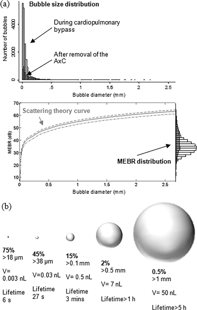
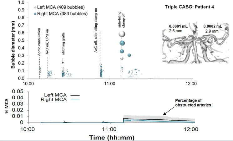
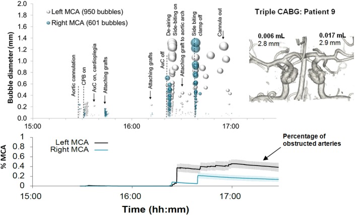
Methods: Transcranial Doppler ultrasound recordings from ten patients undergoing heart surgery were analysed for the presence of embolic signals. The backscattered intensity of each embolic signal was modelled based on ultrasound scattering theory to provide an estimate of bubble diameter. The impact of showers of bubbles on cerebral blood-flow was then investigated using patient-specific Monte-Carlo simulations to model the accumulation and clearance of bubbles within a model vasculature.

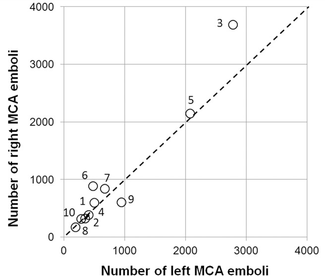
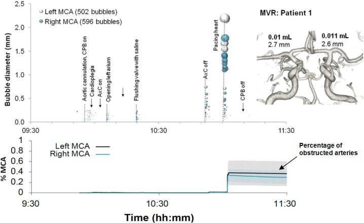
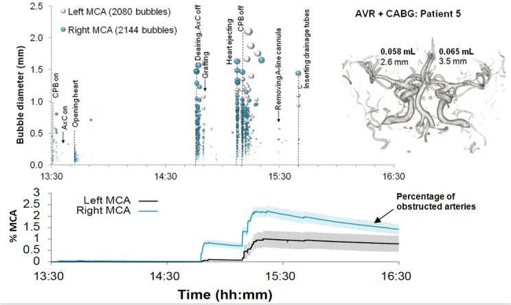
Results: Analysis of Doppler ultrasound recordings revealed a minimum of 371 and maximum of 6476 bubbles entering the middle cerebral artery territories during surgery. This was estimated to correspond to a total volume of air ranging between 0.003 and 0.12 mL. Based on analysis of a total of 18667 embolic signals, the median diameter of bubbles entering the cerebral arteries was 33 μm (IQR: 18 to 69 μm). Although bubble diameters ranged from ~5 μm to 3.5 mm, the majority (85%) were less than 100 μm. Numerous small bubbles detected during cardiopulmonary bypass were estimated by Monte-Carlo simulation to be benign. However, during weaning from bypass, showers containing large macro-bubbles were observed, which were estimated to transiently affect up to 2.2% of arterioles.

Conclusions: Detailed analysis of Doppler ultrasound data can be used to provide an estimate of bubble diameter, total volume of air, and the likely impact of embolic showers on cerebral blood flow. Although bubbles are alarmingly numerous during surgery, our simulations suggest that the majority of bubbles are too small to be harmful.


Conflict of interest statement

Competing Interests: The authors have declared that no competing interests exist.

Figures

Fig 1
Fig 1. Total numbers of bubbles detected in the left and right middle cerebral arteries.
Similar numbers of bubbles were detected in the left and right MCAs of individual patients. Markers are labelled with patient identifiers.
Fig 2
Fig 2. Estimated distribution of bubble sizes detected during cardiac surgery.
(a) Distribution of bubble sizes estimated based on analysis of the ultrasound backscatter (MEBR values) from 18667 embolic signals. Overall, the median diameter of bubbles was 33 μm (IQR: 18 to 69 μm). Signals observed following removal of the aortic cross-clamp are shaded. (b) Percentage of emboli, bubble volume, and predicted dissolve times for 18 μm, 38 μm, 100 μm, 500 μm and 1 mm diameter air bubbles.
Fig 3
Fig 3. Timing and diameters of bubbles in a 60 year old patient during Triple CABG.
Markers denote individual embolic events where the y-axis and marker size indicate estimated bubble diameter. The lower panel displays the predicted number of blocked MCA end arterioles estimated by Monte-Carlo simulation. The shaded regions highlight 95% confidence intervals, based on uncertainty in bubble diameter and variations in outcome between simulations. The inset shows a 3D reconstruction of the circle of Willis labelled with estimated total volume of air and MCA diameters.
Fig 4
Fig 4. Timing and diameters of bubbles in a 76 year old patient during Triple CABG.
Markers denote individual embolic events where the y-axis and marker size indicate estimated bubble diameter. The lower panel displays the predicted number of blocked MCA end arterioles estimated by Monte-Carlo simulation. The shaded regions highlight 95% confidence intervals, based on uncertainty in bubble diameter and variations in outcome between simulations. The inset shows a 3D reconstruction of the circle of Willis labelled with estimated total volume of air and MCA diameters.
Fig 5
Fig 5. Timing and diameters of bubbles in a 71 year old patient during combined MVR and CABG.
Markers denote individual embolic events where the y-axis and marker size indicates estimated bubble diameter. The lower panel displays the predicted number of blocked end arterioles obtained by Monte-Carlo simulation. The shaded regions highlight 95% confidence intervals, based on uncertainty in bubble diameter and variations in outcome between simulations. The inset shows a 3D reconstruction of the circle of Willis labelled with estimated total volume of air and MCA diameters.
Fig 6
Fig 6. Timing and diameters of bubbles in a 55 year old patient during combined AVR and CABG.
Markers denote individual embolic events where the y-axis and marker size indicates estimated bubble diameter. The lower panel displays the predicted number of blocked end arterioles obtained by Monte-Carlo simulation. The shaded regions highlight 95% confidence intervals, based on uncertainty in bubble diameter and variations in outcome between simulations. The inset shows a 3D reconstruction of the circle of Willis labelled with estimated total volume of air and MCA diameters.

References

    1. Barak M, Katz Y (2005) Microbubbles: pathophysiology and clinical implications. Chest 128: 2918–2932. - PubMed
    1. Mitchell S, Gorman D (2002) The pathophysiology of cerebral arterial gas embolism. J Extra Corpor Technol 34: 18–23. - PubMed
    1. Chaudhuri K, Marasco SF (2011) The effect of carbon dioxide insufflation on cognitive function during cardiac surgery. J Card Surg 26: 189–196. 10.1111/j.1540-8191.2011.01217.x - DOI - PubMed
    1. Gerriets T, Schwarz N, Sammer G, Baehr J, Stolz E, et al. (2010) Protecting the brain from gaseous and solid micro-emboli during coronary artery bypass grafting: a randomized controlled trial. Eur Heart J 31: 360–368. 10.1093/eurheartj/ehp178 - DOI - PubMed
    1. Kruis RW, Vlasveld FA, Van Dijk D (2010) The (un)importance of cerebral microemboli. Semin Cardiothorac Vasc Anesth 14: 111–118. 10.1177/1089253210370903 - DOI - PubMed

Publication types