Review
. 2015 Mar 13;112(11):177-84.
doi: 10.3238/arztebl.2015.0177.

Total ankle replacement

Affiliations
Review

Total ankle replacement

Alexej Barg et al. Dtsch Arztebl Int. .

Abstract

Background: About 1% of adults suffer from painful osteoarthritis of the ankle. The current literature contains no information on the percentage of such patients who derive long-term relief of symptoms from conservative treatment. Advanced ankle osteoarthritis can be treated with non-joint-preserving measures, such as total ankle replacement and ankle fusion.

Methods: This review is based on selected relevant publications, guidelines from Germany and abroad, and the authors' personal experience.

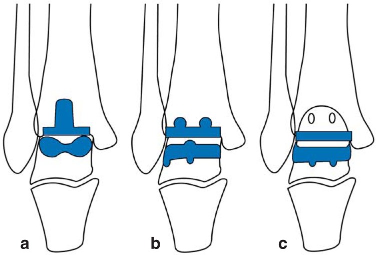
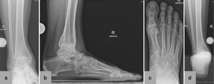
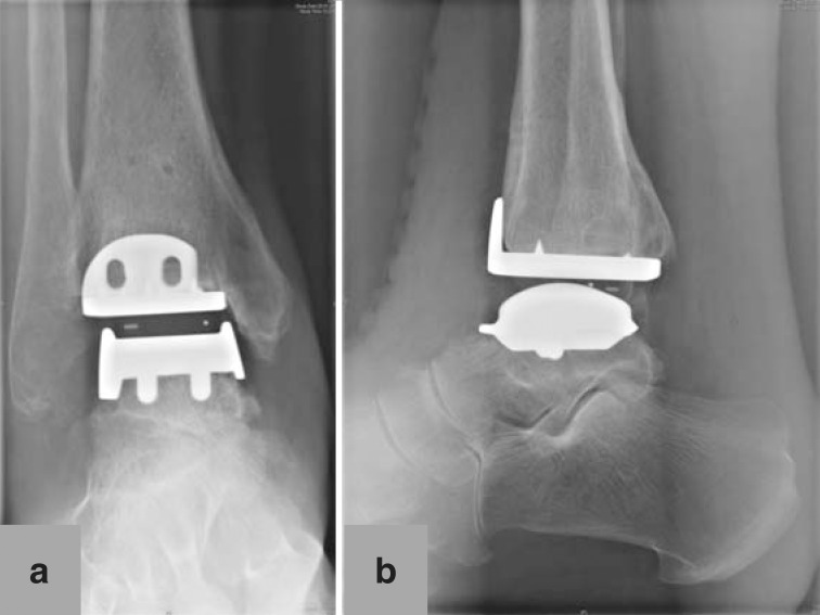
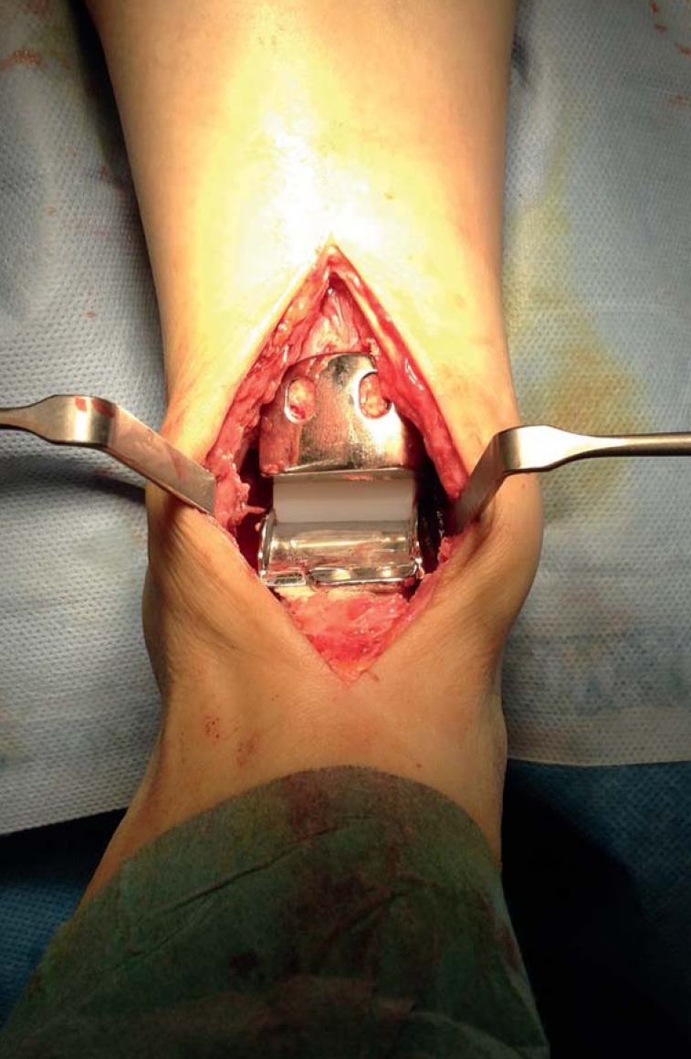
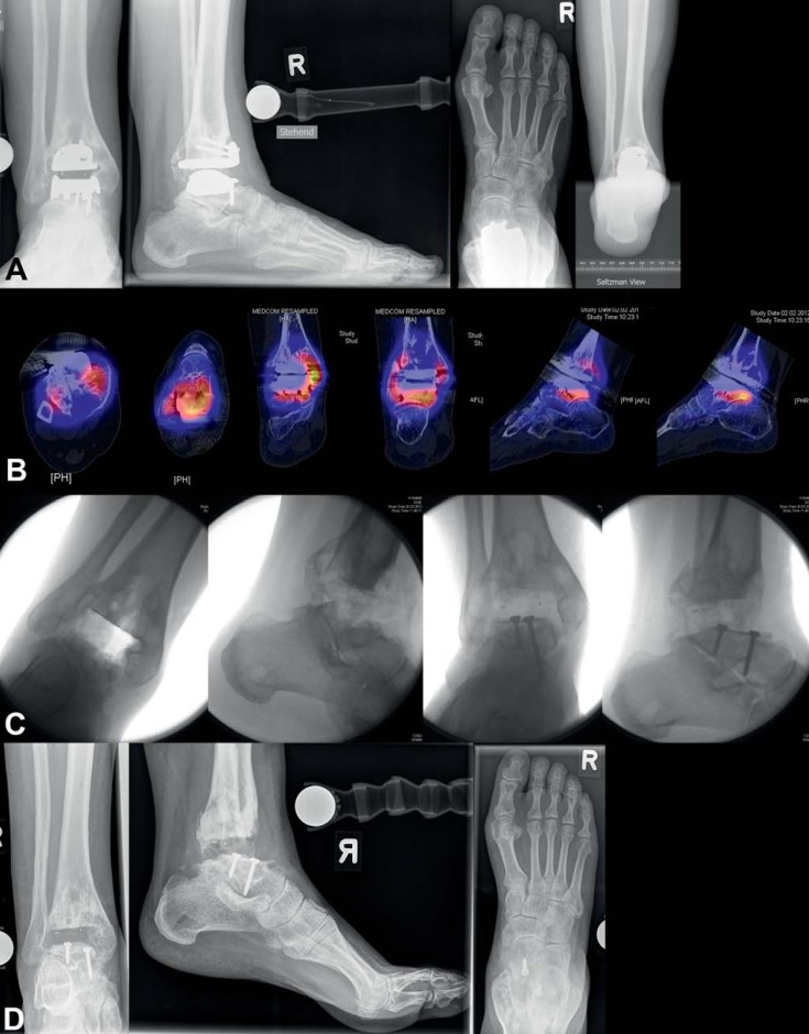
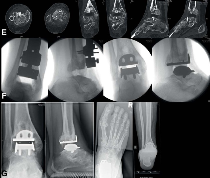
Results: Before surgery is considered, conservative measures such as physiotherapy and orthopedic aids should be used to the fullest possible extent. No randomized trials have yet been published comparing total ankle replacement with ankle fusion. Total ankle replacement with newer types of prosthesis yields good to very good intermediate-term and long-term results, with mean success rates of up to 90% at 10 years (range, 68-100%). Independent risk factors for the failure of ankle replacement are age over 70 years (odds ratio [OR] 3.84), primary osteoarthritis (OR 7.19), post-traumatic osteoarthritis (OR 6.2), and type of prosthesis (e.g., single hydroxyapatite coating: OR 15.04). The average range of motion of the replaced ankle joint is 25° to 30°, with values as high as 60°.

Conclusion: Total ankle replacement is a good treatment option for complete, end-stage ankle arthritis. It can restore joint function and make the patient mobile with little or no pain. There are, however, many contraindications to be taken into account. There is a need for further studies of the biomechanics of arthritic and replaced ankle joints and for long-term follow-up studies of total ankle replacement.


Figures

Figure 1
Figure 1
Modern ankle implant types a) Components with tibial stem b) Components with bars c) Flat components
Figure 2
Figure 2
Preoperative conventional X-ray in standing position of 67-year-old female patient with posttraumatic ankle osteoarthritis following open reduction and internation fixation for trimalleolar luxation fracture 4 years earlier: a) mortise view of ankle; b) lateral view of foot and ankle; c) dorsoplantar view of foot; d) Saltzman view of hindfoot
Figure 3
Figure 3
Postoperative X-ray of 67-year-old female patient 6 weeks after total ankle replacement: a) mortise view of ankle; b) lateral view of ankle
eFigure 1
eFigure 1
Total ankle replacement (HINTEGRA implant) in situ, implanted via anterior approach to ankle. Talar metal components (bottom), polyethylene inlay (white, center), and tibial metal components (top) are visible
eFigure 2
eFigure 2
Aseptic loosening of total ankle replacement 8 years after initial surgery: a) conventional X-rays in standing position show borders of loosening around both implant components, tibial and talar; b) SPECT-CT shows high metabolic activity in the areas around both implant components; c) implant components have been removed, on talar side osseous reconstruction was performed using autograft taken from iliac crest; d) and e) conventional X-rays and CT show good bone consolidation of autograft from iliac crest on the talar side 4 months postoperatively; f) fixation screws on talar side have been removed, HINTEGRA implant (revision components on talar side) were implanted; g) conventional X-rays in standing position show good osseointegration of implant components 6 months postoperatively
eFigure 2
eFigure 2
Aseptic loosening of total ankle replacement 8 years after initial surgery: a) conventional X-rays in standing position show borders of loosening around both implant components, tibial and talar; b) SPECT-CT shows high metabolic activity in the areas around both implant components; c) implant components have been removed, on talar side osseous reconstruction was performed using autograft taken from iliac crest; d) and e) conventional X-rays and CT show good bone consolidation of autograft from iliac crest on the talar side 4 months postoperatively; f) fixation screws on talar side have been removed, HINTEGRA implant (revision components on talar side) were implanted; g) conventional X-rays in standing position show good osseointegration of implant components 6 months postoperatively

References

    1. Barg A, Pagenstert GI, Hugle T, et al. Ankle osteoarthritis: etiology, diagnostics, and classification. Foot Ankle Clin. 2013;18:411–426. - PubMed
    1. Glazebrook M, Daniels T, Younger A, et al. Comparison of health-related quality of life between patients with end-stage ankle and hip arthrosis. J Bone Joint Surg Am. 2008;90:499–505. - PubMed
    1. Saltzman CL, Salamon ML, Blanchard GM, Huff T, Hayes A, Buckwalter JA, et al. Epidemiology of ankle arthritis: report of a consecutive series of 639 patients from a tertiary orthopaedic center. Iowa Orthop J. 2005;25:44–46. - PMC - PubMed
    1. Goost H, Wimmer MD, Barg A, Kabir K, Valderrabano V, Burger C. Fractures of the ankle joint: investigation and treatment options. Dtsch Arztebl Int. 2014;111:377–388. - PMC - PubMed
    1. Valderrabano V, Hintermann B, Horisberger M, Fung TS. Ligamentous posttraumatic ankle osteoarthritis. Am J Sports Med. 2006;34:612–620. - PubMed

MeSH terms