. 2015 Jun 23;43(11):5489-500.
doi: 10.1093/nar/gkv243. Epub 2015 Apr 2.

Next-generation sequencing reveals the biological significance of the N(2),3-ethenoguanine lesion in vivo

Affiliations

Next-generation sequencing reveals the biological significance of the N(2),3-ethenoguanine lesion in vivo

Shiou-chi Chang et al. Nucleic Acids Res. .

Abstract

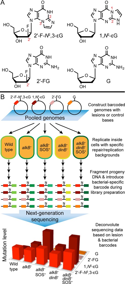
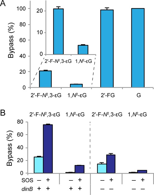
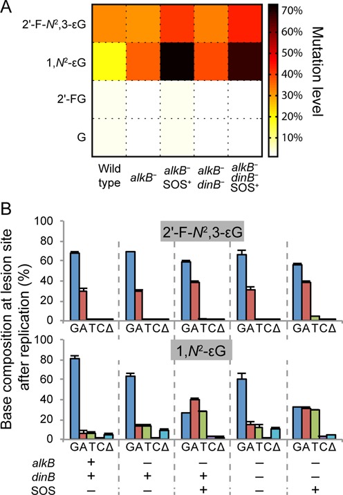
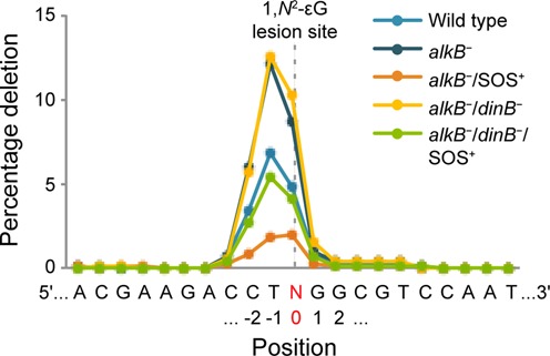
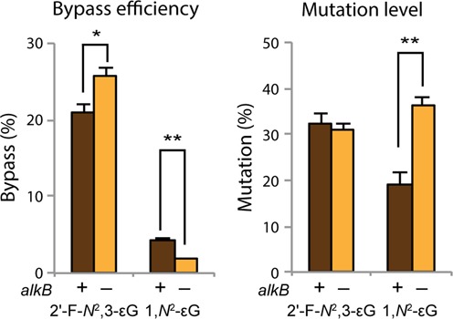
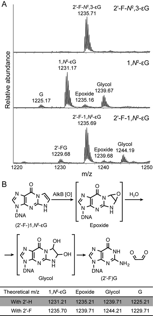
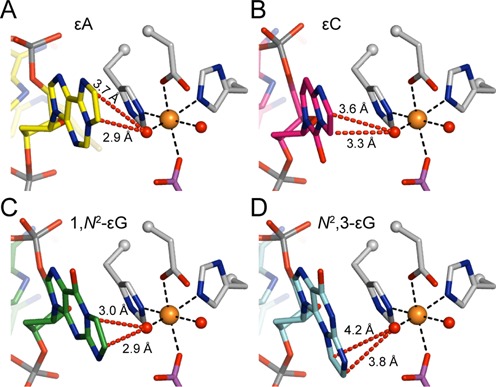
Etheno DNA adducts are a prevalent type of DNA damage caused by vinyl chloride (VC) exposure and oxidative stress. Etheno adducts are mutagenic and may contribute to the initiation of several pathologies; thus, elucidating the pathways by which they induce cellular transformation is critical. Although N(2),3-ethenoguanine (N(2),3-εG) is the most abundant etheno adduct, its biological consequences have not been well characterized in cells due to its labile glycosidic bond. Here, a stabilized 2'-fluoro-2'-deoxyribose analog of N(2),3-εG was used to quantify directly its genotoxicity and mutagenicity. A multiplex method involving next-generation sequencing enabled a large-scale in vivo analysis, in which both N(2),3-εG and its isomer 1,N(2)-ethenoguanine (1,N(2)-εG) were evaluated in various repair and replication backgrounds. We found that N(2),3-εG potently induces G to A transitions, the same mutation previously observed in VC-associated tumors. By contrast, 1,N(2)-εG induces various substitutions and frameshifts. We also found that N(2),3-εG is the only etheno lesion that cannot be repaired by AlkB, which partially explains its persistence. Both εG lesions are strong replication blocks and DinB, a translesion polymerase, facilitates the mutagenic bypass of both lesions. Collectively, our results indicate that N(2),3-εG is a biologically important lesion and may have a functional role in VC-induced or inflammation-driven carcinogenesis.


Figures

Figure 1.
Figure 1.
Experimental overview. (A) Structures of the modified DNA bases and controls (shown as deoxynucleosides) investigated for genotoxic and mutagenic properties. Numbers in red show the key atom positions on the nucleosides. (B) Schematic representation of the in vivo mutagenesis assay with next-generation sequencing. M13 single-stranded vectors, each containing a site-specific lesion and a lesion-specific barcode sequence, were mixed in a known ratio and introduced into cells with specific repair and replication backgrounds. After in vivo replication, progeny DNA from each repair/replication background was isolated, amplified and fragmented to generate sequencing libraries. N represents the site, in progeny, that had originally contained the lesion, and the colored box to the left of N symbolizes the lesion-specific barcode (Barcode 1). A second set of barcodes (Barcode 2, to the right of N), designating the repair/replication backgrounds and biological replicates were also introduced at the library preparation step. The resulting DNA was pooled and subjected to next-generation sequencing. The genotoxicity and mutagenicity of each lesion under each bacterial condition were determined from the sequencing data, which were sorted according to the two sets of barcodes.
Figure 2.
Figure 2.
Replicative bypass efficiencies of the εG lesions. Lower bypass efficiency indicates higher lesion genotoxicity. (A) Bypass efficiencies of 2′-F-N2,3-εG and 1,N2-εG lesions, as well as 2′-FG and G controls in wild type (HK81) cells, with error bars representing one standard deviation (SD) (N = 3). Inset shows zoomed-in details of the 2′-F-N2,3-εG and 1,N2-εG results. (B) Bypass efficiencies of the εG lesions with or without SOS induction and in the presence or absence of DinB. The results shown in this figure (also tabulated in Supplementary Table S2) were obtained in an alkB background. Error bars represent one SD (N = 3).
Figure 3.
Figure 3.
Lesion mutation frequencies. (A) A heat map representation of the mutation frequencies of 2′-F-N2,3-εG, 1,N2-εG, 2′-FG and G in all five repair/replication backgrounds investigated. (B) Mutation frequency and specificity of 2′-F-N2,3-εG and 1,N2-εG under the five bacterial conditions investigated, with error bars representing one SD (N = 3). G, A, T and C indicate the possible bases present at site N (Figure 1) in progeny genomes (i.e. the base present at the lesion site after in vivo replication), and Δ denotes the occurrence of deletions at the lesion site after replication. Results presented in this figure are also tabulated in Supplementary Table S4.
Figure 4.
Figure 4.
The frequency of deletions induced by 1,N2-εG at the lesion site and adjacent positions. Each line represents the averaged results from the three independent biological replicates of a specific repair and replication background. N, highlighted in red, denotes the lesion site (position 0).
Figure 5.
Figure 5.
Replicative bypass efficiencies and mutation levels of 2′-F-N2,3-εG and 1,N2-εG in the presence and absence of AlkB. Error bars represent one SD (N = 3). Statistical significance was determined by a two-tailed, heteroscedastic, Student's t-test (*P < 0.05, **P < 0.002).
Figure 6.
Figure 6.
Susceptibility of εG lesions to AlkB repair in vitro. (A) Mass spectra of 16-mer oligonucleotides containing either a 2′-F-N2,3-εG, 1,N2-εG or 2′-F-1,N2-εG following 1 h incubation with AlkB. Data represent the −4 charge envelopes and the observed monoisotopic peak value is labeled above each peak envelope. (B) The proposed mechanism of AlkB repair on (2′-F-)1,N2-εG. Theoretical m/z values of the −4 charged ions of the 16-mer oligonucleotides starting materials, putative reaction intermediates and repair products are listed for both the 2′-deoxy (2′-H) and 2′-fluoro (2′-F) versions of the bases. A recent theoretical study proposed that repair of εA by AlkB may be mediated by a zwitterionic species, and that some of the species observed by the MS analysis could be byproducts rather than repair intermediates (56). However, the ability of AlkB to repair the εG lesions is not affected by this hypothesis.
Figure 7.
Figure 7.
Models of the four etheno lesions in the active site of AlkB. (A) AlkB active site with an εA lesion (yellow carbons, PDB ID: 3O1P), a known good substrate for AlkB, and with the iron(IV)-oxo intermediate modeled (see below). (BD) Models of AlkB with (B) εC (pink carbons), (C) 1,N2-εG (green carbons), and (D) N2,3-εG (cyan carbons) lesions in the active site. Models of 1,N2-εG and N2,3-εG are based on the crystal structure of εA in AlkB (PDB ID: 3O1P) and the model of εC is based on the crystal structure of 3-methylcytosine (3mC) in AlkB (PDB ID: 3O1M, Supplementary Figure S6). In all panels, selected AlkB amino acid residues are shown in grey, iron-bound succinate in purple, the iron ion as an orange sphere, and iron-bound oxygens (or water molecules) as red spheres. The iron(IV)-oxo intermediate is modeled based on a recent study, with an iron-oxygen distance of 1.62 Å (56). Distances from the iron(IV)-oxo oxygen atom to the exocyclic etheno carbon atoms are shown as red dashed lines.

Similar articles

Cited by

References

    1. Breslow L., Hoaglin L., Rasmussen G., Abrams H.K. Occupations and cigarette smoking as factors in lung cancer. Am. J. Public Health Nations Health. 1954;44:171–181. - PMC - PubMed
    1. Abdulla F.R., Feldman S.R., Williford P.M., Krowchuk D., Kaur M. Tanning and skin cancer. Pediatr. Dermatol. 2005;22:501–512. - PubMed
    1. Wogan G.N. Dietary factors and special epidemiological situations of liver cancer in Thailand and Africa. Cancer Res. 1975;35:3499–3502. - PubMed
    1. Triantafillidis J.K., Nasioulas G., Kosmidis P.A. Colorectal cancer and inflammatory bowel disease: epidemiology, risk factors, mechanisms of carcinogenesis and prevention strategies. Anticancer Res. 2009;29:2727–2737. - PubMed
    1. International Agency for Research on Cancer. IARC Monogr. Eval. Carcinog. Risks Hum. Vol. 97. Lyon: International Agency for Research on Cancer; 2008. pp. 311–443. - PMC - PubMed

Publication types

LinkOut - more resources