

Amplitude-integrated EEG in newborns with critical congenital heart disease predicts preoperative brain magnetic resonance imaging findings
- PMID: 25838043
- PMCID: PMC4442075
- DOI: 10.1016/j.pediatrneurol.2015.02.026
Amplitude-integrated EEG in newborns with critical congenital heart disease predicts preoperative brain magnetic resonance imaging findings
Abstract
Objective: The study aims are to evaluate cerebral background patterns using amplitude-integrated electroencephalography in newborns with critical congenital heart disease, determine if amplitude-integrated electroencephalography is predictive of preoperative brain injury, and assess the incidence of preoperative seizures. We hypothesize that amplitude-integrated electroencephalography will show abnormal background patterns in the early preoperative period in infants with congenital heart disease that have preoperative brain injury on magnetic resonance imaging.
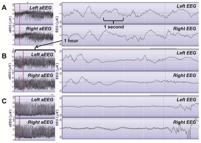
Methods: Twenty-four newborns with congenital heart disease requiring surgery at younger than 30 days of age were prospectively enrolled within the first 3 days of age at a tertiary care pediatric hospital. Infants had amplitude-integrated electroencephalography for 24 hours beginning close to birth and preoperative brain magnetic resonance imaging. The amplitude-integrated electroencephalographies were read to determine if the background pattern was normal, mildly abnormal, or severely abnormal. The presence of seizures and sleep-wake cycling were noted. The preoperative brain magnetic resonance imaging scans were used for brain injury and brain atrophy assessment.
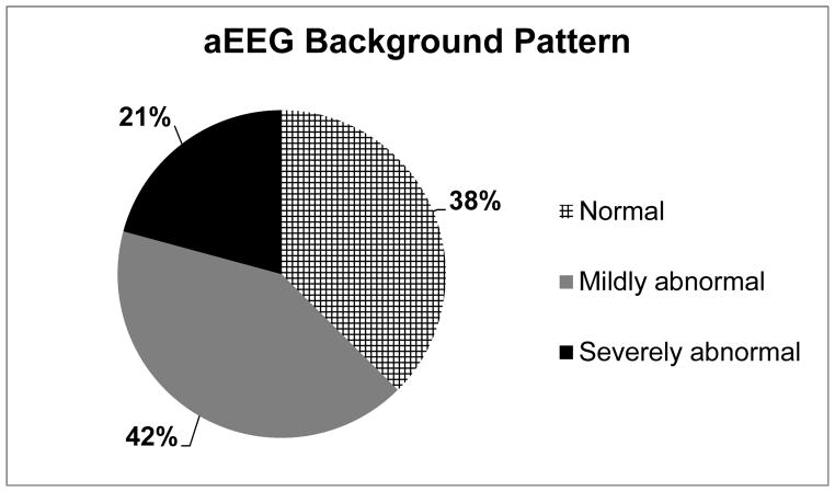
Results: Fifteen of 24 infants had abnormal amplitude-integrated electroencephalography at 0.71 (0-2) (mean [range]) days of age. In five infants, the background pattern was severely abnormal. (burst suppression and/or continuous low voltage). Of the 15 infants with abnormal amplitude-integrated electroencephalography, 9 (60%) had brain injury. One infant with brain injury had a seizure on amplitude-integrated electroencephalography. A severely abnormal background pattern on amplitude-integrated electroencephalography was associated with brain atrophy (P = 0.03) and absent sleep-wake cycling (P = 0.022).
Conclusion: Background cerebral activity is abnormal on amplitude-integrated electroencephalography following birth in newborns with congenital heart disease who have findings of brain injury and/or brain atrophy on preoperative brain magnetic resonance imaging.
Keywords: brain injury; congenital heart disease; electroencephalography; neuromonitoring; newborn; seizures.
Copyright © 2015 Elsevier Inc. All rights reserved.
Conflict of interest statement
Figures
References
-
- Mahle WT, Tavani F, Zimmerman RA, Nicolson SC, Galli KK, Gaynor JW, et al. An MRI study of neurological injury before and after congenital heart surgery. Circulation. 2002;106(12 Suppl 1):I109–14. - PubMed
-
- McQuillen PS, Barkovich AJ, Hamrick SE, Perez M, Ward P, Glidden DV, et al. Temporal and anatomic risk profile of brain injury with neonatal repair of congenital heart defects. Stroke. 2007;38(2 Suppl):736–41. - PubMed
-
- Shellhaas RA, Chang T, Tsuchida T, Scher MS, Riviello JJ, Abend NS, et al. The American Clinical Neurophysiology Society’s Guideline on Continuous Electroencephalography Monitoring in Neonates. J Clin Neurophysiol. 2011;28:611–7. - PubMed
-
- de Vries LS, Toet MC. Amplitude integrated electroencephalography in the full-term newborn. Clin Perinatol. 2006;33:619–32. - PubMed
Publication types
MeSH terms
Grants and funding
LinkOut - more resources
Full Text Sources
Other Literature Sources
Medical
