Review
. 2015 May 14;125(20):3049-58.
doi: 10.1182/blood-2014-11-568881. Epub 2015 Apr 2.

Pathogenesis beyond the cancer clone(s) in multiple myeloma

Affiliations
Review

Pathogenesis beyond the cancer clone(s) in multiple myeloma

Giada Bianchi et al. Blood. .

Abstract

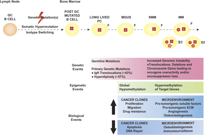
Over the past 4 decades, basic research has provided crucial information regarding the cellular and molecular biology of cancer. In particular, the relevance of cancer microenvironment (including both cellular and noncellular elements) and the concept of clonal evolution and heterogeneity have emerged as important in cancer pathogenesis, immunologic escape, and resistance to therapy. Multiple myeloma (MM), a cancer of terminally differentiated plasma cells, is emblematic of the impact of cancer microenvironment and the role of clonal evolution. Although genetic and epigenetic aberrations occur in MM and evolve over time under the pressure of exogenous stimuli, they are also largely present in premalignant plasma cell dyscrasia such as monoclonal gammopathy of undetermined significance (MGUS) and smoldering multiple myeloma (SMM), suggesting that genetic mutations alone are necessary, but not sufficient, for myeloma transformation. The role of bone marrow microenvironment in mediating survival, proliferation, and resistance to therapy in myeloma is well established; and although an appealing speculation, its role in fostering the evolution of MGUS or SMM into MM is yet to be proven. In this review, we discuss MM pathogenesis with a particular emphasis on the role of bone marrow microenvironment.


Figures

Figure 1
Figure 1
Pathogenesis of MM. The orange round cell represents a normal B cell, whereas the yellow round cell is a mutated, post–germinal center (GC) B lymphocyte that later differentiates into a long-lived PC (yellow oval). In MM pathogenesis, the initial genetic event (red square) is thought to occur in the GC, facilitated by the processes of somatic hypermutation and isotype switching, and characterizes the founder clone (F). Later genetic mutations occur at the time of transformation to MM (red circle), with de novo mutations (red geometric shapes) acquired during disease evolution and heterogeneously present in different subclones (S1 and S2). The genetic, epigenetic, and biological events occurring in the cancer clones and BM microenvironment during the evolution of premalignant dyscrasia to MM are outlined in the pink, green, and blue boxes, respectively. ECM, extracellular matrix.
Figure 2
Figure 2
Role of the BM niche in MM pathogenesis. The blue oval in the center is the MMC, with its close interplay with cellular and acellular components of the BM. The pale orange ovals represent relevant cytokines/chemokines in the BM milieu. Dotted arrows indicate differentiation, whereas solid arrows indicate secretion and/or effect on a target cell. Yellow squares contain a synopsis of the overall effect of cytokines and cell-to-cell contact on the target cell. Key signaling cascades, transmembrane proteins, and intracellular organelles, which are of interest for molecularly targeted therapies, are represented. BMSC, BM stromal cell; CAF, cancer-associated fibroblast; CCL2, chemokine (C-C motif) ligand 2; CTLA4, cytotoxic T-lymphocyte-associated protein 4; CXCL12, chemokine (C-X-C motif) ligand 12; CXCR4, chemokine (C-X-C motif) receptor 4; DKK-1, dickkopf WNT signaling pathway inhibitor 1; ER, endoplasmic reticulum; FN, fibronectin; HGF, hepatocyte growth factor; HIF-1α, hypoxia-inducible factor 1α; ICAM1, intercellular adhesion molecule 1; IGF-1, insulinlike growth factor 1; ITGB1, integrin β1; ITGB2, integrin β2; JNK, c-JUN N-terminal kinase; MAPK1, mitogen-activated protein kinase 1; MDSC, myeloid-derived suppressor cell; MIP-1α, macrophage inflammatory protein 1α; MUC-1, mucin 1; NK-T cells, natural killer T cells; OPG, osteoprotegerin; PD-1, programmed cell death 1; PD-L1, programmed ligand death 1; RANK, receptor activator of NF-κB; RANKL, RANK ligand; RBC, red blood cell; TGF-β, transforming growth factor β; TH17, T helper 17 cell; Treg, regulatory T cell; VCAM1, vascular cell adhesion molecule 1; VEGFA, vascular endothelial growth factor A; WNT, wingless-type. Adapted from Bianchi and Anderson with permission.

References

    1. Anderson KC, Carrasco RD. Pathogenesis of myeloma. Annu Rev Pathol. 2011;6:249–274. - PubMed
    1. Hauser AE, Muehlinghaus G, Manz RA, et al. Long-lived plasma cells in immunity and inflammation. Ann N Y Acad Sci. 2003;987:266–269. - PubMed
    1. Seifert M, Scholtysik R, Küppers R. Origin and pathogenesis of B cell lymphomas. Methods Mol Biol. 2013;971:1–25. - PubMed
    1. Morgan GJ, Walker BA, Davies FE. The genetic architecture of multiple myeloma. Nat Rev Cancer. 2012;12(5):335–348. - PubMed
    1. Kuehl WM, Bergsagel PL. Molecular pathogenesis of multiple myeloma and its premalignant precursor. J Clin Invest. 2012;122(10):3456–3463. - PMC - PubMed

Publication types