Review
. 2015 Apr 3;348(6230):62-8.
doi: 10.1126/science.aaa4967.

Adoptive cell transfer as personalized immunotherapy for human cancer

Affiliations
Review

Adoptive cell transfer as personalized immunotherapy for human cancer

Steven A Rosenberg et al. Science. .

Abstract

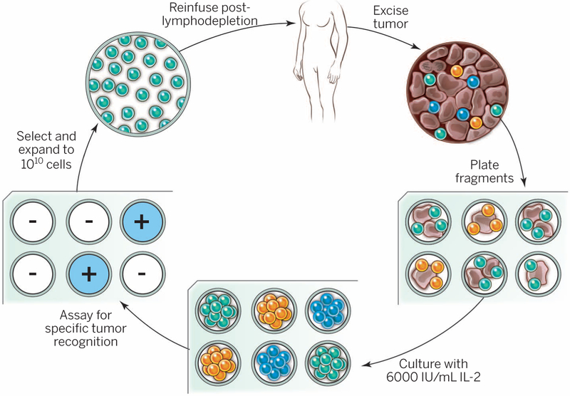
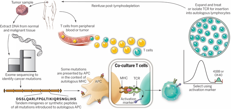
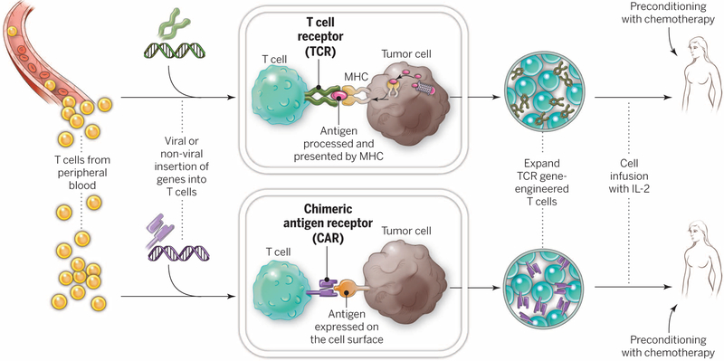
Adoptive cell therapy (ACT) is a highly personalized cancer therapy that involves administration to the cancer-bearing host of immune cells with direct anticancer activity. ACT using naturally occurring tumor-reactive lymphocytes has mediated durable, complete regressions in patients with melanoma, probably by targeting somatic mutations exclusive to each cancer. These results have expanded the reach of ACT to the treatment of common epithelial cancers. In addition, the ability to genetically engineer lymphocytes to express conventional T cell receptors or chimeric antigen receptors has further extended the successful application of ACT for cancer treatment.


Figures

Fig. 1.
Fig. 1.. General schema for using the adoptive cell transfer of naturally occurring autologous TILs.
The resected melanoma specimen is digested into a single-cell suspension or divided into multiple tumor fragments that are individually grown in IL-2. Lymphocytes overgrow, destroy tumors within 2 to 3 weeks, and generate pure cultures of lymphocytes that can be tested for reactivity in coculture assays. Individual cultures are then rapidly expanded in the presence of excess irradiated feeder lymphocytes, OKT3, and IL-2. By approximately 5 to 6 weeks after resecting the tumor, up to 1011 lymphocytes can be obtained for infusion into patients.
Fig. 2.
Fig. 2.. A substantial increase in cell persistence and the incidence and duration of clinical responses is observed when patients received a lymphodepleting preparative regimen before the cell infusion.
The most frequently used lymphodepleting preparative regimen consists of 60 mg/kg cyclophosphamide given for 2 days and 25 mg/m2 fludarabine administered over 5 days, followed by T cells and IL-2 administration.
Fig. 3.
Fig. 3.. A “blueprint” for the treatment of patients with T cells recognizing tumor-specific mutations.
The sequences of exomic DNA from tumor cells and normal cells from the same patient are compared to identify tumor-specific mutations. Knowledge of these mutations can then be used to synthesize either minigenes or polypeptides encoding each mutated amino acid flanked by 10 to 12 amino acids. These peptides or minigenes can be expressed by a patient’s autologous APCs, where they are processed and presented in the context of a patient’s MHC. Coculture of the patient’s T cells with these APCs can be used to identify all mutations processed and presented in the context of all of a patient’s MHC class I and class II molecules. The identification of individual mutations responsible for tumor recognition is possible because T cells express activation markers, such as 41BB (CD8+ T cells) and OX40 (CD4+ T cells), when they recognize their cognate target antigen. T cells expressing the activation marker can then by purified using flow cytometry before their expansion and reinfusion into the tumor-bearing patient.
Fig. 4.
Fig. 4.. Gene-modification of peripheral blood lymphocytes.
In an attempt to broaden the reach of ACT to other cancers, techniques are being developed to introduce antitumor receptors into normal T cells that could be used for therapy. The top panel shows the insertion of a conventional TCR into a patient’s T lymphocytes, followed by the expansion and infusion back into the patient. The bottom panel shows the insertion of a CAR into a patient’s T cell, followed by the expansion of these cells and their re-infusion. TCRs and CARs are fundamentally different in their structures and in the structures that they recognize. TCRs are composed of one a chain and one β chain, and they recognize antigens that have been processed and presented by one of the patient’s own MHC molecules. CARs are artificial receptors that can be constructed by linking the variable regions of the antibody heavy and light chains to intracellular signaling chains (such as CD3-zeta, CD28, 41BB) alone or in combination with other signaling moieties. CARs recognize antigens that do not need to be MHC-restricted, but they must be presented on the tumor cell surface.

References

    1. Delorme EJ, Alexander P, Lancet 284, 117–120 (1964). - PubMed
    1. Fefer A, Cancer Res. 29, 2177–2183 (1969). - PubMed
    1. Fernandez-Cruz E, Woda BA, Feldman JD, J. Exp. Med 152, 823–841 (1980). - PMC - PubMed
    1. Berendt MJ, North RJ, J. Exp. Med 151, 69–80 (1980). - PMC - PubMed
    1. Morgan DA, Ruscetti FW, Gallo R, Science 193, 1007–1008 (1976). - PubMed

Publication types

Substances