Case Reports
. 2015 Mar;14(Suppl 1):300-7.
doi: 10.1007/s12663-013-0507-6. Epub 2013 Apr 2.

Maxillo-Mandibular Cemento-ossifying Fibroma: A Rare Case Report

Affiliations
Case Reports

Maxillo-Mandibular Cemento-ossifying Fibroma: A Rare Case Report

Ranjit Kumar Peravali et al. J Maxillofac Oral Surg. 2015 Mar.

Abstract

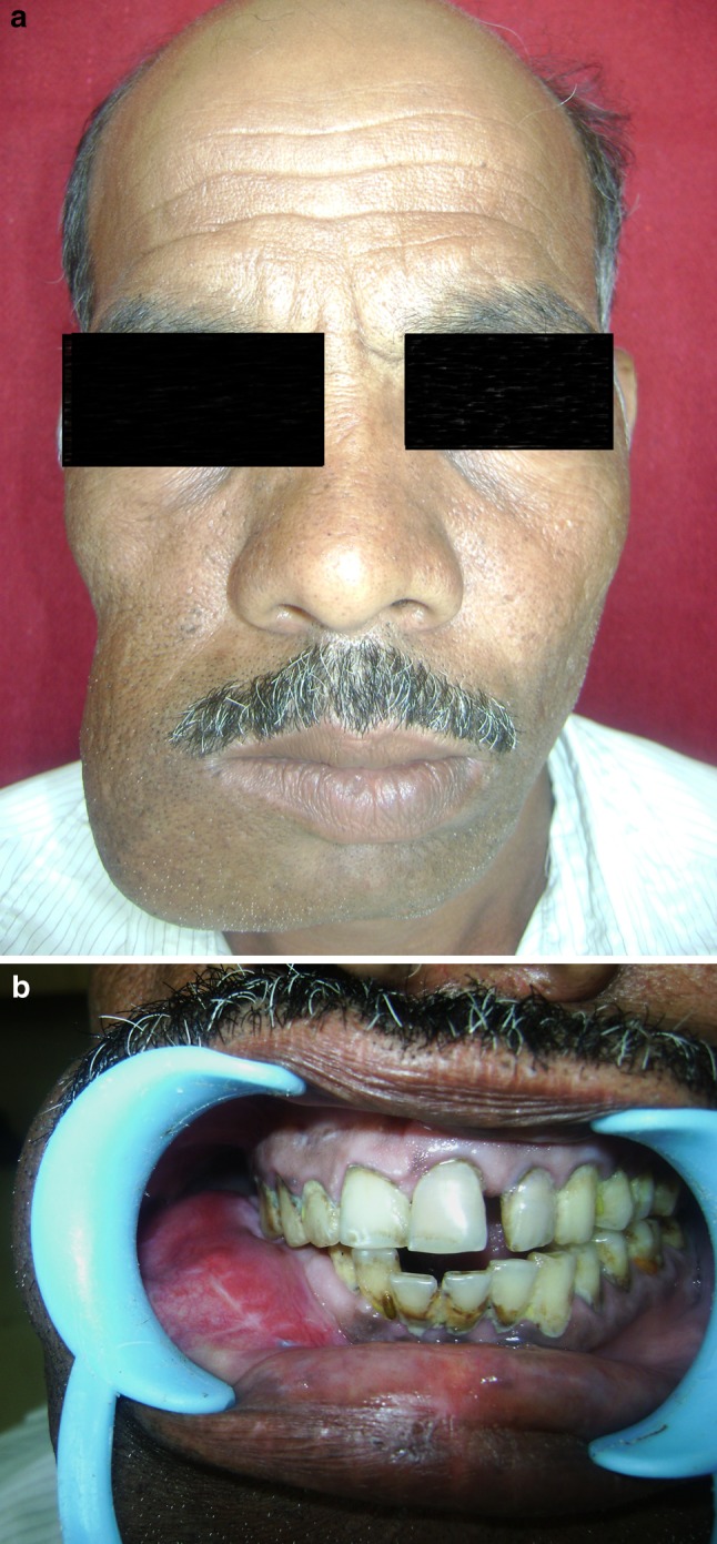
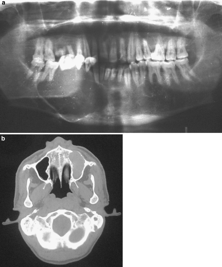
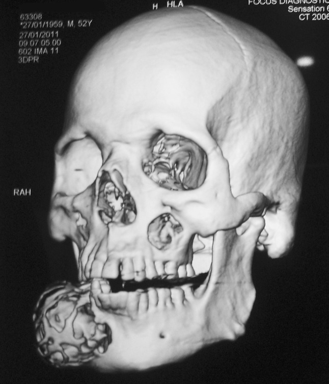
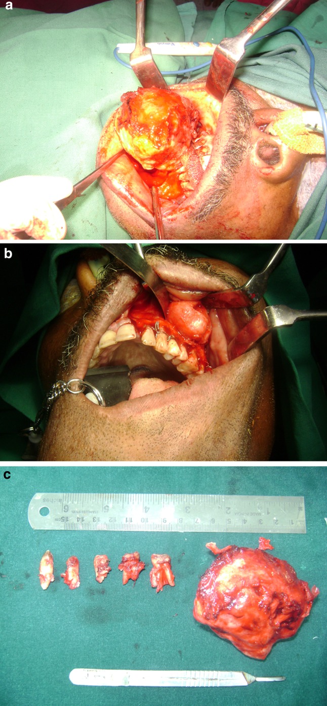
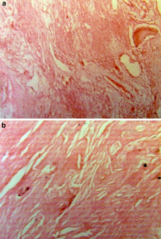
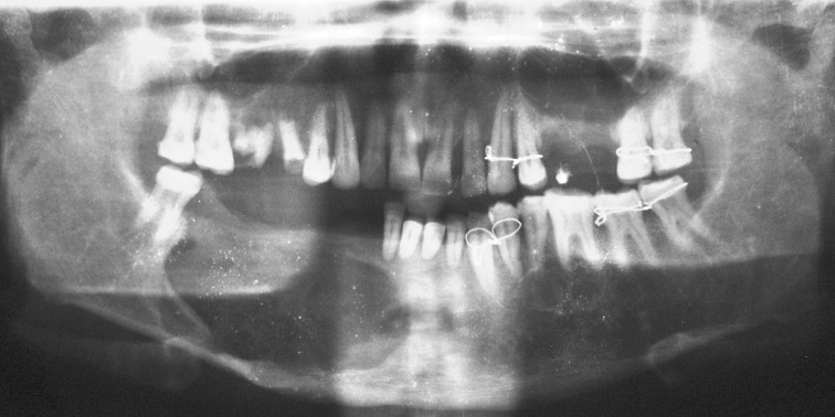
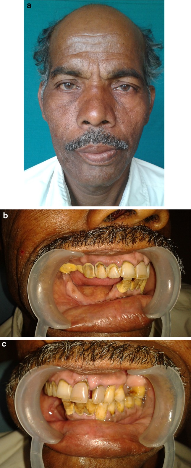
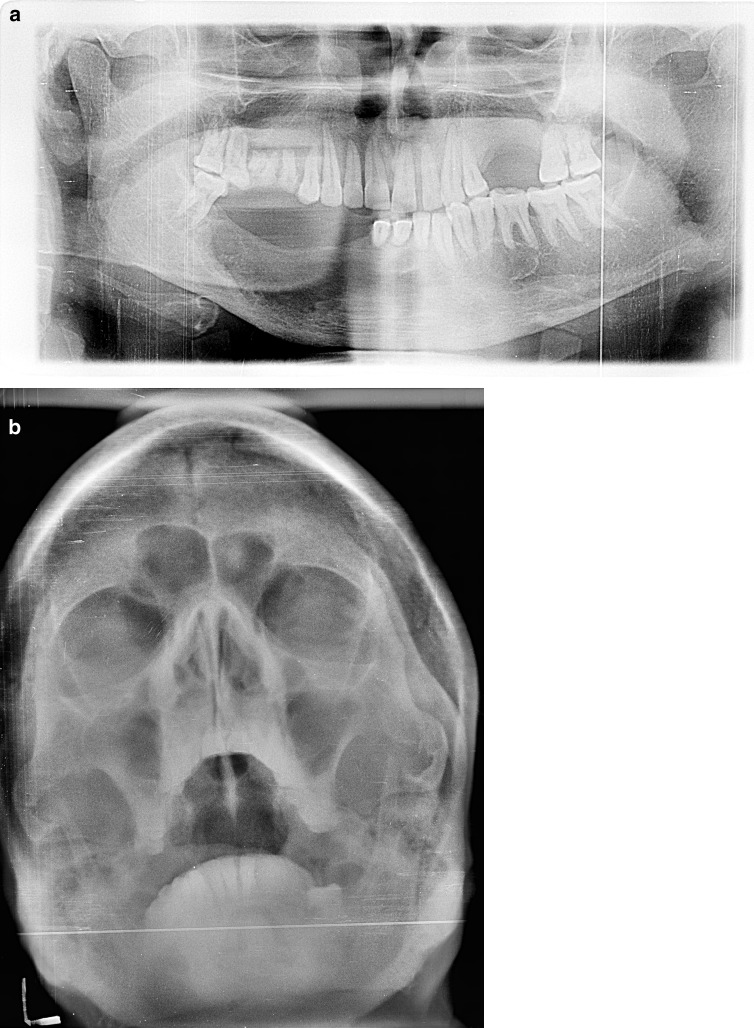
Cemento-ossifying fibroma (COF) is a benign fibro osseous lesion of the jaws which has been described as a demarcated or rarely encapsulated neoplasm consisting of fibrous tissue and varying amounts of mineralized material resembling bone and/or cementum (Dinkar et al. in IJDA 2(4):45-47, 2010). Majority of lesions occur in the mandible and only few cases of COFs of the maxillary sinus and bilateral COFs of the mandible have been reported in literature (Dinkar et al. in IJDA 2(4):45-47, 2010; Tamiolakis et al. in Acta Stomatol Croat 39(3):319-321, 2005; Hamner et al. in Oral Surg Oral Med Oral Pathol 26(4):579-587, 1968; Gunaseelan et al. in Oral Med Oral Pathol Oral Radiol Endod 104:e21-e25, 2007). These lesions have a very low recurrence rate (Ertug et al. in Quintessence Int 35(10):808-810, 2004) and are generally treated by enucleation. In this paper we present a rare case of COF occurring in both the maxilla and mandible of the same patient. Only one such case (Takeda and Fujioka in Int J Oral Maxillofac Surg 16(3):368-371, 1987) has been reported in literature so far.

Keywords: Bi-jaw; Cemento-ossifying fibroma; Maxilla-mandibular.


Figures

Fig. 1
Fig. 1
a Marked facial asymmetry seen with a diffuse swelling in the right body of the mandible region. b Bucco-lingual cortical expansion causing vestibular obliteration, extending from 42 to 47 with intact overlying mucosa. The teeth were lingually displaced, deranging the occlusion
Fig. 2
Fig. 2
a OPG showing a well-defined radiolucency with sclerotic margins extending from lower right canine to distal surface of second molar. Thinning out of the lower border of mandible and displacement of teeth is evident. b CT axial section showing a hyperdense lesion within the left maxillary antrum
Fig. 3
Fig. 3
3D reconstruction of the CT image showing extent of the lesion
Fig. 4
Fig. 4
a Lesion in the right body of the mandible being excised in-toto. b Lesion in the left maxillary antrum. c Resected mandibular lesion along with extracted teeth
Fig. 5
Fig. 5
H&E stained tissue section of mandibular lesion (a) and antral lesion (b) showing dense connective tissue stroma with thick collagen bundles and numerous endothelial lined blood vessels with haphazardly arranged fibroblasts. Basophilic calcified masses are seen, exhibiting osteoblastic rimming
Fig. 6
Fig. 6
Immediate post operative orthopantomograph
Fig. 7
Fig. 7
a One year post-operative photograph in frontal profile. b One year post-operative intra oral photograph showing no evidence of recurrence of the mandibular lesion. c One year post-operative intra oral photograph showing no evidence of recurrence of the maxillary lesion
Fig. 8
Fig. 8
a One year post-operative orthopantomograph showing no recurrence of the mandibular lesion. b One year post-operative PNS view showing no recurrence of the maxillary lesion

References

    1. Dinkar D, Arathi K, Ahmed S, Rai N. Bilateral cemento ossifying fibroma of mandible. IJDA. 2010;2:45–47.
    1. Tamiolakis D, Thomaidis V, Tsamis I. Cemento-ossifying fibroma of the maxilla: a case report. Acta Stomatol Croat. 2005;39:319–321.
    1. Hamner JE, Lightbody PM, Ketcham AS, Swerdlow S, Bethesda (1968) Cemento-ossifying fibroma of the maxilla. Oral Surg Oral Med Oral Pathol 26:579–587 - PubMed
    1. Gunaseelan R, Parameswaran A, Erumbala R, Kannan R. Large cemento-ossifying fibroma of the maxilla causing proptosis: a case report. Oral Med Oral Pathol Oral Radiol Endod. 2007;104:e21–e25. doi: 10.1016/j.tripleo.2007.04.026. - DOI - PubMed
    1. Ertug E, Meral VG, Saysel M. Cemento-ossifying fibroma: a case report. Quintessence Int. 2004;35:808–810. - PubMed

Publication types

LinkOut - more resources