

Primary intraosseous hydatid cyst of femur
- PMID: 25838934
- PMCID: PMC4376981
- DOI: 10.5812/ircmj.21070
Primary intraosseous hydatid cyst of femur
Abstract
Introduction: Echinococcosis is a parasitic and zoonotic disease of animals and humans. The cause is Echinococcus granulosus and occasionally, Echinococcus multilocularis. Hydatid cysts are mostly seen in the liver and lungs, although almost all organs and systemscan be involvement. Hydatid cysts seen with bone involvement comprise approximately 3% of all hydatid cysts. Even if a long period of survey is possible, it is still difficult to eradicate the disease and effect a cure.
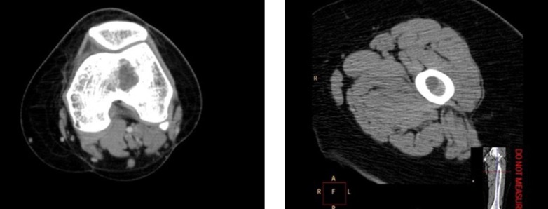
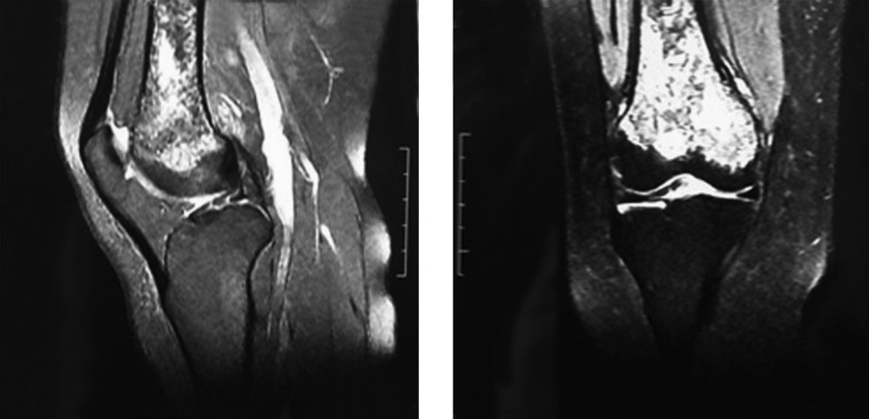
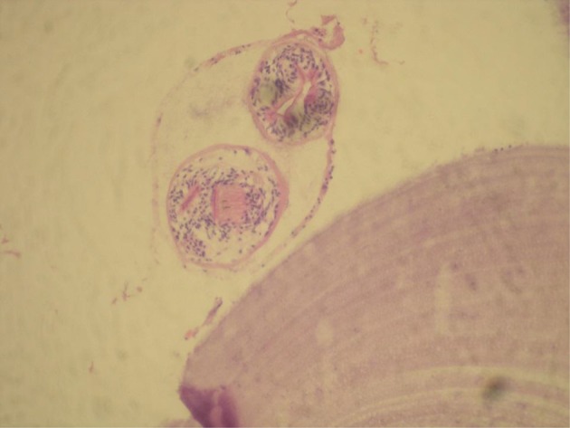
Case presentation: In this study, an evaluation was made of a patient referred at Yozgat State Hospital Orthopedics and Traumatology Polyclinic with complaints of pain in her left thigh close to the knee. After examinations of plain radiographs, computerized tomography, magnetic resonance images, and blood parameters, a diagnosis was made of left femoral intramedullary hydatid cyst from excised intraoperative material. Throughout a 6-month follow-up period, there was no recurrence and functional results were good.
Conclusions: Based on this report (of a patient presented with an intramedullary cyst in the long bones), the primary bone hydatid cyst disease should be kept in mind and be investigated in the differential diagnosis.
Keywords: Cyst; Echinococcus granulosus; Femur; Hydatid.
Figures
References
-
- Hepgul G, Tihan D, Kocael P, Dogan Y, Ozturk T, Cihan A. [Case report: primary splenic hydatidosis]. Turkiye Parazitol Derg. 2010;34(3):184–6. - PubMed
-
- Natarajan MV, Kumar AK, Sivaseelam A, Iyakutty P, Raja M, Rajagopal TS. Using a custom mega prosthesis to treat hydatidosis of bone: a report of 3 cases. J Orthop Surg (Hong Kong). 2002;10(2):203–5. - PubMed
-
- Zlitni M, Ezzaouia K, Lebib H, Karray M, Kooli M, Mestiri M. Hydatid cyst of bone: diagnosis and treatment. World J Surg. 2001;25(1):75–82. - PubMed
-
- Canale ST, Beaty JH. In: Campbells Operative Orthopaedics. Başbozkurt M, Yıldız C, editors. İstanbul: Sun medical book house; 2011.
Publication types
LinkOut - more resources
Full Text Sources
Other Literature Sources