. 2015 Apr 3;3(1-2):e982426.
doi: 10.4161/21688370.2014.982426. eCollection 2015.

Roles and regulation of the mucus barrier in the gut

Affiliations

Roles and regulation of the mucus barrier in the gut

Steve Cornick et al. Tissue Barriers. .

Abstract

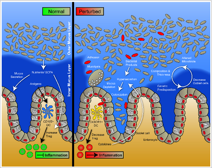
The gastrointestinal tract is coated by a thick layer of mucus that forms the front line of innate host defense. Mucus consists of high molecular weight glycoproteins called mucins that are synthesized and secreted by goblet cells and functions primarily to lubricate the epithelium and protect it from damage by noxious substances. Recent studies have also suggested the involvement of goblet cells and mucins in complex immune functions such as antigen presentation and tolerance. Under normal physiological conditions, goblet cells continually produce mucins to replenish and maintain the mucus barrier; however, goblet cell function can be disrupted by various factors that can affect the integrity of the mucus barrier. Some of these factors such as microbes, microbial toxins and cytokines can stimulate or inhibit mucin production and secretion, alter the chemical composition of mucins or degrade the mucus layer. This can lead to a compromised mucus barrier and subsequently to various pathological conditions like chronic inflammatory diseases. Insight into how these factors modulate the mucus barrier in the gut is necessary in order to develop strategies to combat these disorders.

Keywords: Barrier function; CD, Crohns disease; ER stress; ERAD, ER-associated protein degradation; EhCP5, Entamoeba histolytica cysteine protease 5; FAS, fatty acid synthase; GI, gastrointestinal; GalNAc, N-Acetylgalactosamine; Goblet cell; IBD; IBD, Inflammatory bowel disease; Innate defense; LLO, Listeriolysin O; LPS, Lipopolysaccharide; MUC2; MucBP, Mucin binding proteins; Mucin; SCFA, short chain fatty acids; Secretory response; UC, Ulcerative colitis; UPR, unfolded protein response; Unfolded protein response.


Figures

Figure 1.
Figure 1.
The mucus layer under normal and perturbed conditions. The gut mucosa is covered by a thick layer of mucus that acts as a protective barrier against harmful substances. The barrier consists of 2 layers, an inner mucus layer and an outer mucus layer. Commensal and environmental microbes colonize and remain in the outer mucus layer by binding to mucus via lectin-like molecules, while the inner mucus layer is relatively sterile. Mucus is composed of high molecular weight glycoproteins called mucins. Mucins are produced and packaged into secretory granules within the goblet cell cytoplasm and secreted at the apical membrane. Under normal conditions, goblet cells continuously produce mucus to maintain a functional mucus layer; however, genetic and environmental factors can modulate goblet cell function and mucus production. Short chain fatty acids (SCFAs) produced by commensal microorganisms not only provide nutrients for colonocytes but also increase mucus production and secretion. Furthermore, SCFAs also modulate immune homeostasis and tolerance in the intestines. Under perturbed conditions during infection, pathogens can induce mucin hypersecretion, inhibit mucus production, degrade mucus or induce changes in mucin glycosylation that can lead to disruption of the mucus barrier. Altered barrier function can subsequently cause commensal and pathogenic microbes as well as microbial products to translocate to the epithelial surface. This in turn can trigger immune cell response and cytokine production leading to inflammation.
Figure 2.
Figure 2.
Endoplasmic reticulum (ER) stress and unfolded protein response (UPR) in goblet cells. Various factors can induce ER stress in goblet cells, which can lead to a reduction in mucin synthesis and a diminished mucus layer. During ER stress, the UPR is activated to restore ER homeostasis. Glucose-regulating peptide 78 (GRP78) dissociates from the UPR initiating molecules and binds misfolded proteins. [PKR (double-stranded RNA-dependent protein kinase)-like ER kinase] (PERK), upon dissociation from GRP78 becomes active and phosphorylates eukaryotic initiation factor-2α (eIF2α) leading to inhibition of mRNA translation. This reduces the protein load trafficking to the ER. The mRNA encoding the transcription factor activating transcription factor 4 (ATF4) is however translated. ATF4 then moves to the nucleus to up-regulate UPR genes. On becoming active, inositol-requiring enzyme 1 activates Xbox binding protein (XBP1). Activated XBP1 then translocate to the nucleus and up-regulates UPR target genes. Activating transcription factor 6 (ATF6) moves to the Golgi apparatus after dissociation from GRP78, where it is cleaved by proteases to form an active transcription factor, which moves to the nucleus to modulate UPR gene expression. ER stress causes misfolding of mucins in the ER. Furthermore, inhibition of mRNA synthesis by the PERK arm can diminish mucin translation. The overall effect is that, decreased synthesis of mucins can lead to fewer mucin granules and a thinner mucus layer.

References

    1. McCracken VJ, Lorenz RG. The gastrointestinal ecosystem: a precarious alliance among epithelium, immunity and microbiota. Cell Microbiol 2001; 3:1-11; PMID:; http://dx.doi.org/10.1046/j.1462-5822.2001.00090.x - DOI - PubMed
    1. Hollingsworth MA, Swanson BJ. Mucins in cancer: protection and control of the cell surface. Nat Rev Cancer 2004; 4:45-60; PMID:; http://dx.doi.org/10.1038/nrc1251 - DOI - PubMed
    1. Boltin D, Perets TT, Vilkin A, Niv Y. Mucin function in inflammatory bowel disease, an update. J Clin Gastroenterol 2013; 47:106-11; PMID:; http://dx.doi.org/10.1097/MCG.0b013e3182688e73 - DOI - PubMed
    1. Forstner G. Signal transduction, packaging and secretion of mucins. Annu Rev Physiol 1995; 57:585-605; PMID:; http://dx.doi.org/10.1146/annurev.ph.57.030195.003101 - DOI - PubMed
    1. Phillips TE, Phillips TH, Neutra MR. Regulation of intestinal goblet cell secretion. III. Isolated intestinal epithelium. Am J Physiol 1984; 247:G674-G681; PMID: - PubMed

LinkOut - more resources