. 2015:2015:627923.
doi: 10.1155/2015/627923. Epub 2015 Mar 9.

Dexamethasone enhanced functional recovery after sciatic nerve crush injury in rats

Affiliations

Dexamethasone enhanced functional recovery after sciatic nerve crush injury in rats

Xinhong Feng et al. Biomed Res Int. 2015.

Abstract

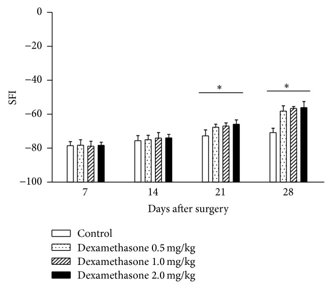
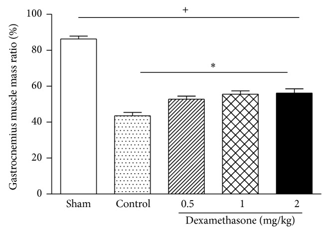
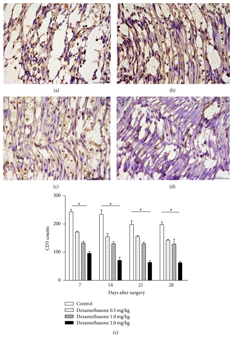
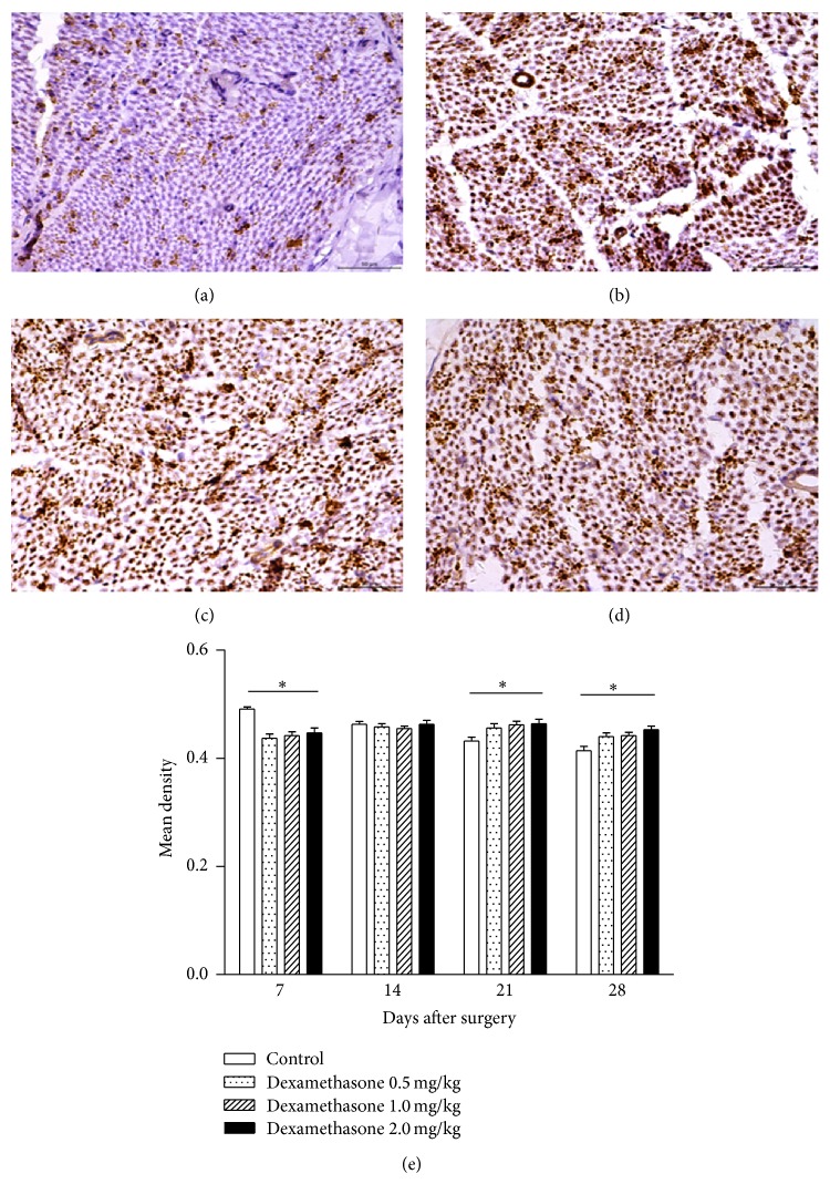
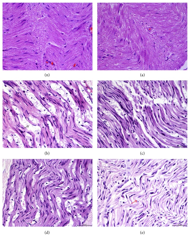
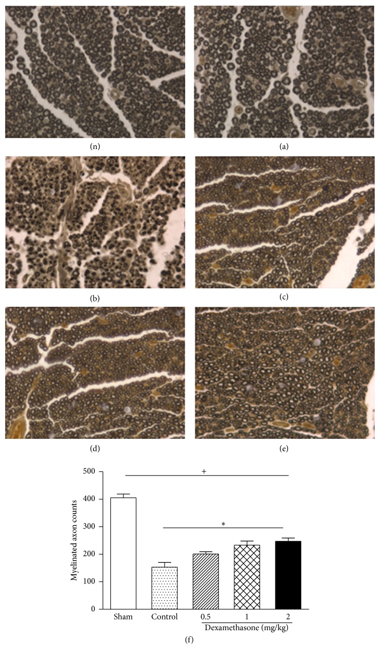
Dexamethasone is currently used for the treatment of peripheral nerve injury, but its mechanisms of action are not completely understood. Inflammation/immune response at the site of nerve lesion is known to be an essential trigger of the pathological changes that have a critical impact on nerve repair and regeneration. In this study, we observed the effects of various doses of dexamethasone on the functional recovery after sciatic nerve crush injury in a rat model. Motor functional recovery was monitored by walking track analysis and gastrocnemius muscle mass ratio. The myelinated axon number was counted by morphometric analysis. Rats administered dexamethasone by local intramuscular injection had a higher nerve function index value, increased gastrocnemius muscle mass ratio, reduced Wallerian degeneration severity, and enhanced regenerated myelinated nerve fibers. Immunohistochemical analysis was performed for CD3 expression, which is a marker for T-cell activation, and infiltration in the sciatic nerve. Dexamethasone-injected rats had fewer CD3-positive cells compared to controls. Furthermore, we found increased expression of GAP-43, which is a factor associated with development and plasticity of the nervous system, in rat nerves receiving dexamethasone. These results provide strong evidence that dexamethasone enhances sciatic nerve regeneration and function recovery in a rat model of sciatic nerve injury through immunosuppressive and potential neurotrophic effects.


Figures

Figure 1
Figure 1
Effects of dexamethasone on the sciatic function index (SFI) 7, 14, 21, and 28 days after surgery. Rats were administered local intramuscular injections with 0.5, 1, or 2 mg/kg dexamethasone, or saline once daily after surgery. The values are represented as means ± SD for 6 rats per group. *Significant difference compared to control, P < 0.05.
Figure 2
Figure 2
Effects of dexamethasone on the gastrocnemius muscle ratio 28 days after surgery. Rats were administered local intramuscular injections with 0.5, 1, or 2 mg/kg dexamethasone, or saline once daily after surgery. The values are represented as means ± SD for 6 rats per group. +Significant difference compared to sham, P < 0.01. *Significant difference compared to control, P < 0.05.
Figure 3
Figure 3
Effects of dexamethasone on T-cell infiltration. ((a)–(d)) Immunohistochemical staining of nerve tissue on day 28 after surgery (magnification, 400x). (a) Control group; ((b)–(d)) 0.5, 1, and 2 mg/kg dexamethasone-treated groups, respectively. Data in the sham group are not shown; (e) CD3-positive cell counts at each time point after surgery. The values represent means ± SD for 6 rats per group. *Significant difference compared to control, P < 0.01.
Figure 4
Figure 4
Effects of dexamethasone on mean density of GAP-43. ((a)–(d)) Immunohistochemical staining of nerve tissue on day 28 after surgery (magnification, 400x). (a) Control group; ((b)–(d)) 0.5, 1, and 2 mg/kg dexamethasone-treated groups, respectively. Data in the sham group are not shown. (e) Mean density of GAP-43 at each time point after surgery. The values represent means ± SD for 6 rats per group. *Significant difference compared to control, P < 0.05.
Figure 5
Figure 5
Histological analysis by H&E staining of rat sciatic nerves on day 28 after surgery. (n) Sciatic nerve on nonoperated side. Group (a) (sham) shows well-organized myelin sheets, round axons, and absence of infiltrating cells. Group (b) shows several areas of edema and degraded myelin sheets (black arrow) and several infiltrating mononuclear cells. Groups (c)–(e) show effects of dexamethasone on the injured sciatic nerves. The red arrow shows that alleviated areas of edema and nerve fibers seem to be better organized than those in group (b). Magnification, ×400.
Figure 6
Figure 6
Effects of dexamethasone on the myelinated axon counts on day 28 after surgery. ((n), (a)–(e)) Histological micrographs of nerve tissue stained with osmium tetroxide staining (magnification 400x). (n) shows the sciatic nerve on nonoperated side; (a) sham group; (b) control group; ((c)–(e)) 0.5, 1, and 2 mg/kg dexamethasone-treated groups, respectively. (f) Total myelinated axon counts. Rats were administered local intramuscular injections with 0.5, 1, or 2 mg/kg dexamethasone, or saline once a day after surgery. The values represent means ± SD for 6 rats per group. +Significant difference compared to sham, P < 0.01. *Significant difference compared to control, P < 0.05.

References

    1. Kim T.-H., Yoon S.-J., Lee W.-C., et al. Protective effect of GCSB-5, an herbal preparation, against peripheral nerve injury in rats. Journal of Ethnopharmacology. 2011;136(2):297–304. doi: 10.1016/j.jep.2011.04.037. - DOI - PubMed
    1. Burnett M. G., Zager E. L. Pathophysiology of peripheral nerve injury: a brief review. Neurosurg Focus. 2004;16, article E1 - PubMed
    1. Steinman L. Elaborate interactions between the immune and nervous systems. Nature Immunology. 2004;5(6):575–581. doi: 10.1038/ni1078. - DOI - PubMed
    1. Watkins L. R., Hutchinson M. R., Milligan E. D., Maier S. F. ‘Listening’ and ‘talking’ to neurons: implications of immune activation for pain control and increasing the efficacy of opioids. Brain Research Reviews. 2007;56(1):148–169. doi: 10.1016/j.brainresrev.2007.06.006. - DOI - PMC - PubMed
    1. Salegio E. A. A., Pollard A. N., Smith M., Zhou X.-F. Sciatic nerve conditioning lesion increases macrophage response but it does not promote the regeneration of injured optic nerves. Brain Research. 2010;1361:12–22. doi: 10.1016/j.brainres.2010.09.015. - DOI - PubMed

MeSH terms

LinkOut - more resources