. 2015 Apr 30;6(12):9908-23.
doi: 10.18632/oncotarget.3532.

Interleukin-21 sustains inflammatory signals that contribute to sporadic colon tumorigenesis

Affiliations

Interleukin-21 sustains inflammatory signals that contribute to sporadic colon tumorigenesis

Veronica De Simone et al. Oncotarget. .

Abstract

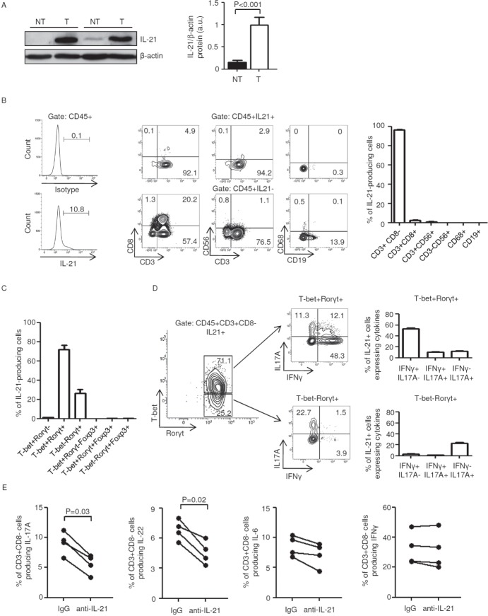
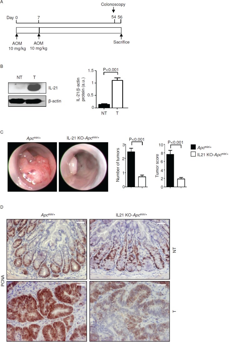
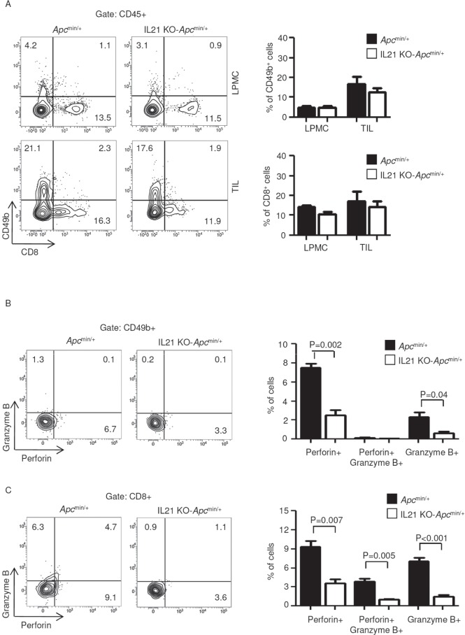
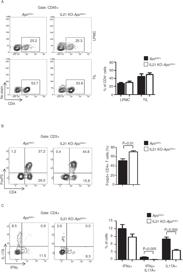
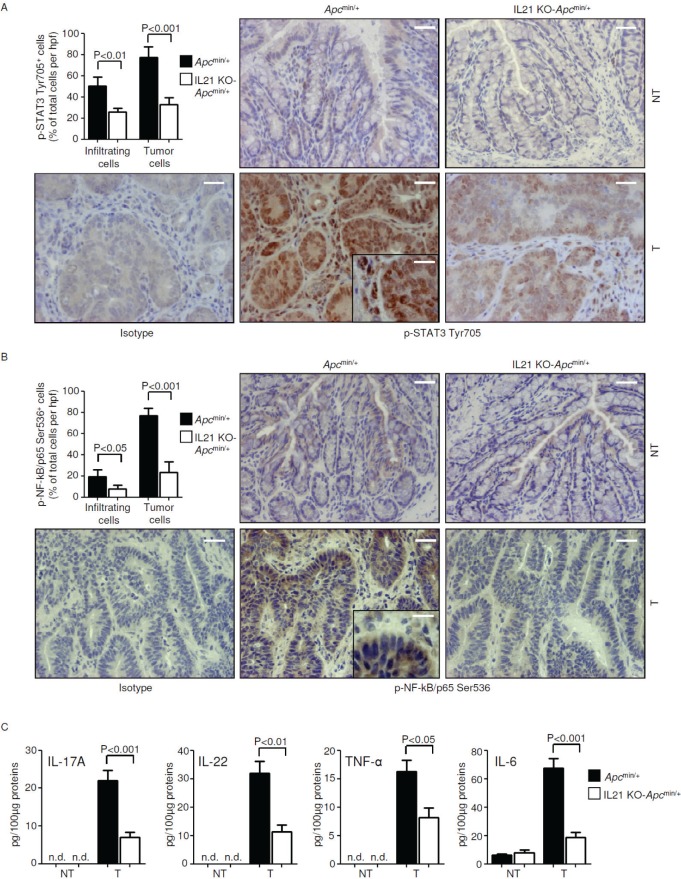
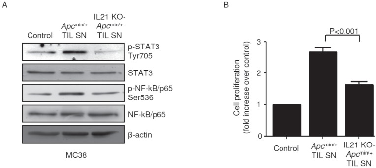
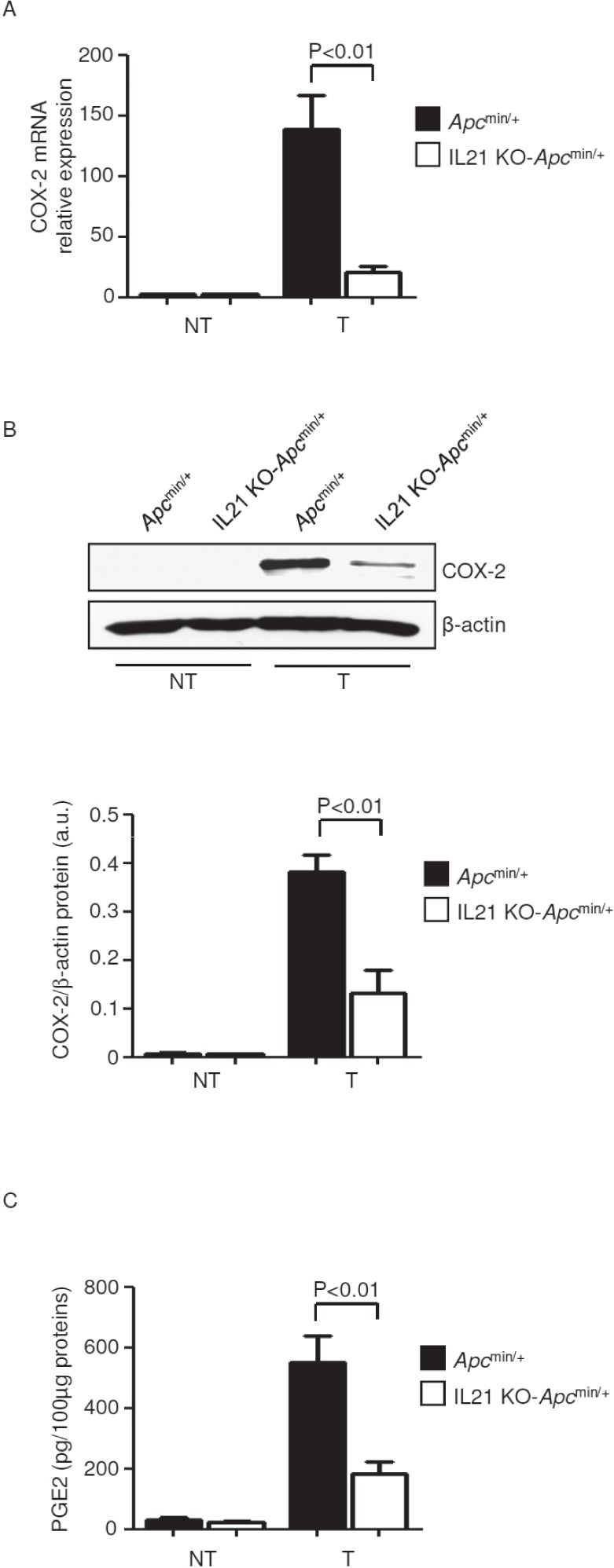
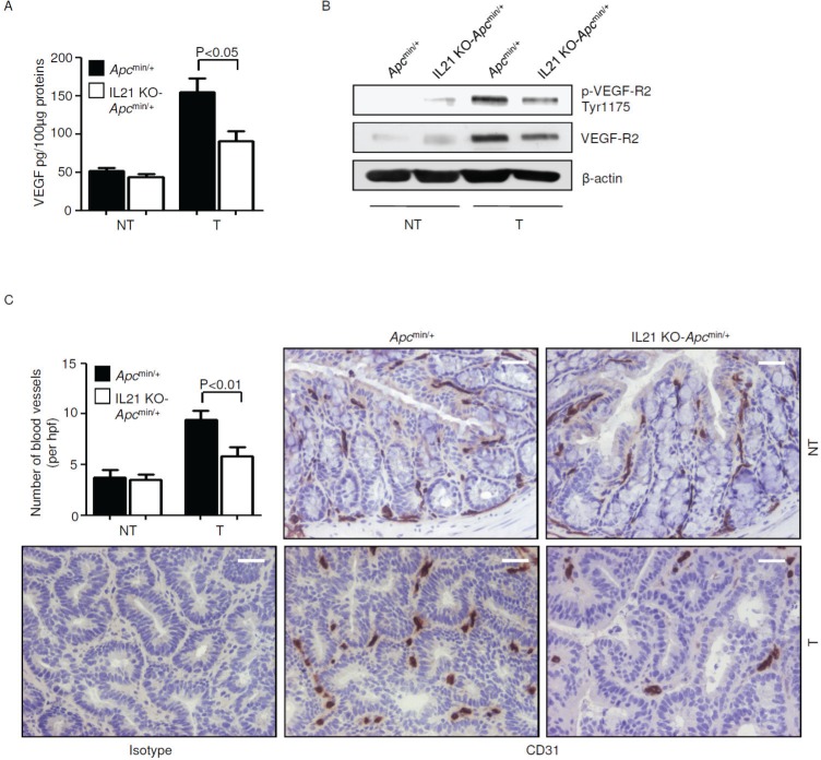
Interleukin (IL)-21 triggers inflammatory signals that contribute to the growth of neoplastic cells in mouse models of colitis-associated colorectal cancer (CRC). Because most CRCs are sporadic and arise in the absence of overt inflammation we have investigated the role of IL-21 in these tumors in mouse and man. IL-21 was highly expressed in human sporadic CRC and produced mostly by IFN-γ-expressing T-bet/RORγt double-positive CD3+CD8- cells. Stimulation of human CRC cell lines with IL-21 did not directly activate the oncogenic transcription factors STAT3 and NF-kB and did not affect CRC cell proliferation and survival. In contrast, IL-21 modulated the production of protumorigenic factors by human tumor infiltrating T cells. IL-21 was upregulated in the neoplastic areas, as compared with non-tumor mucosa, of Apc(min/+) mice, and genetic ablation of IL-21 in such mice resulted in a marked decrease of both tumor incidence and size. IL-21 deficiency was associated with reduced STAT3/NF-kB activation in both immune cells and neoplastic cells, diminished synthesis of protumorigenic cytokines (that is, IL-17A, IL-22, TNF-α and IL-6), downregulation of COX-2/PGE2 pathway and decreased angiogenesis in the lesions of Apc(min/+) mice. Altogether, data suggest that IL-21 promotes a protumorigenic inflammatory circuit that ultimately sustains the development of sporadic CRC.

Keywords: Apcmin/+ mice; COX-2/PGE2; STAT3; VEGF.


Conflict of interest statement

CONFLICTs OF INTEREST

The authors declare no conflict of interest.

Figures

Figure 1
Figure 1. Characterization of IL-21-producing cells in human sporadic colorectal cancer (CRC)
A. Total proteins extracted from both tumor (T) and non-tumor (NT) areas of two patients with sporadic CRC were evaluated for IL-21 expression by western blotting. β-actin was used as loading control. The figure is representative of five separate experiments. Right inset. Quantitative analysis of IL-21/β-actin protein ratio. Values are expressed in arbitrary units (a.u.) and indicate the mean ± SEM of all experiments (n=10). B. IL-21 is preferentially made by CD3+CD8− cells in tumor infiltrating leukocytes (TILs). Left panel. Representative histograms showing the percentage of CD45+IL-21+ cells in TILs. The example is representative of five experiments in which cells isolated from five patients with sporadic CRC were analyzed. Numbers above lines indicate the percentages of CD45+IL-21+ cells. Staining of cells with PE-conjugated control isotype IgG is also shown. Central panels. Representative dot-plots showing the fraction of IL-21+ cells in TILs. CD45+IL-21+ and CD45+IL-21- cells were gated and then analyzed for the indicated markers. The numbers indicate the percentage of cells in the designated quadrants. The example is representative of five experiments in which cells isolated from five patients with sporadic CRC were analyzed. Right panel. Representative histograms showing the fraction of IL-21+ cells in TILs. CD45+IL-21+ cells were gated and then analyzed for the indicated markers. Data are expressed as mean ± SEM of five experiments in which cells isolated from five patients with sporadic CRC were analyzed. C-D. IL-21 is preferentially made by IFN-γ-expressing T-bet/RORγt double-positive CD3+CD8− cells in human sporadic CRC. C. Representative histograms showing the percentages of IL-21-producing CD3+CD8− cells expressing the indicated transcription factors. Data are expressed as mean ± SEM of five experiments in which TILs isolated from five patients with sporadic CRC were analyzed. D. IL-17A and IFN-γ expression was analyzed in T-bet-RORγt+ and T-bet/RORγt double-positive IL-21-producing CD3+CD8− cells by flow cytometry. The numbers indicate the percentage of cells in the designated quadrants. Right panels. Percentages of T-bet-RORγt+ and T-bet/RORγt double-positive IL-21-producing CD3+CD8− cells co-expressing IL-17A and/or IFN-γ. Data are expressed as mean ± SEM of five experiments in which TILs isolated from five patients with sporadic CRC were analyzed. E. TILs isolated from four patients with sporadic CRC were cultured either with a control IgG or anti-IL-21 antibody (both used at 10 μg/ml) for 48 h. Each point represents the percentage of IL-17A-, IL-22-, IL-6- and IFN-γ-producing CD45+CD3+CD8− cells, assessed by flow cytometry, in TILs isolated from a single patient.
Figure 2
Figure 2. IL-21 deficiency significantly reduces colon tumorigenesis in Apcmin/+ mice
A. Experimental protocol used to assess the effect of IL-21 ablation on colon tumorigenesis in Apcmin/+ mice. B. Representative western blotting showing IL-21 expression in colon tissues taken from Apcmin/+ mice killed on day 56. β-actin was used as loading control. One of five representative experiments is shown. NT, non-tumor area, T, tumor area. Right inset. Quantitative analysis of IL-21/β-actin protein ratio in total extracts of T and NT colon tissues taken from Apcmin/+ mice killed on day 56 as measured by densitometry scanning of western blots. Values are expressed in arbitrary units (a.u.) and indicate the mean ± SEM of all experiments (n=5). C. Representative endoscopic pictures of colon tumors developed in Apcmin/+ mice and IL-21 KO-Apcmin/+ mice. Graphs show the number of lesions and the endoscopic scoring of tumors. Data indicate mean ± SEM of four independent experiments in which at least four mice per group were considered. D. Representative images showing PCNA immunostaining in colon sections taken from Apcmin/+ mice and IL-21 KO-Apcmin/+ mice killed on day 56. The scale bars are 20μm. One of four representative experiments in which similar results were obtained is shown.
Figure 3
Figure 3. IL-21 deficiency in Apcmin/+ mice associates with a reduced fraction of perforin and/or granzyme B-producing CD49b- and CD8−positive cells in TILs
A. Representative dot-plots showing CD49b- and CD8−positive cells in LPMCs and TILs isolated from the colon of Apcmin/+ mice and IL-21 KO-Apcmin/+ mice killed on day 56. Numbers indicate the percentages of cells in the designated quadrants. One of four representative experiments is shown. Right insets. Representative histograms showing the fraction of CD49b- and CD8−positive cells in LPMCs and TILs isolated from the colon of Apcmin/+ mice and IL-21 KO-Apcmin/+ mice killed on day 56. CD45-positive cells were gated and analyzed for the indicated markers. Values are mean ± SEM of two independent experiments containing at least two mice per group. B-C. TILs isolated from the colon of Apcmin/+ mice and IL-21 KO-Apcmin/+ mice killed on day 56 were assessed for perforin- and granzyme B-expressing CD49b- (B) and CD8−positive cells (C) by flow cytometry. The numbers indicate the percentage of cells in the designated quadrants. Right panels. Percentages of CD49b- (B) and CD8−positive cells (C) co-expressing perforin and/or granzyme B. Data are expressed as mean ± SEM of two independent experiments containing at least two mice per group.
Figure 4
Figure 4. IL-21 deficiency in Apcmin/+ mice associates with a functional switch of TIL-derived CD4-positive cells
A. Representative dot-plots showing CD4-positive cells in LPMCs and TILs isolated from the colon of Apcmin/+ mice and IL-21 KO-Apcmin/+ mice killed on day 56. Numbers indicate the percentages of cells in the designated quadrants. One of four representative experiments is shown. Right insets. Representative histograms showing the fraction of CD4-positive cells in LPMCs and TILs isolated from the colon of Apcmin/+ mice and IL-21 KO-Apcmin/+ mice killed on day 56. CD45-positive cells were gated and analyzed for the indicated markers. Values are mean ± SEM of two independent experiments containing at least two mice per group. B. TILs isolated from the colon of Apcmin/+ mice and IL-21 KO-Apcmin/+ mice killed on day 56 were assessed for Foxp3-expressing CD4-positive cells by flow cytometry. The numbers indicate the percentage of cells in the designated quadrants. Graph shows the fraction of Foxp3-positive CD4 T cells in TILs isolated from the colon of Apcmin/+ mice and IL-21 KO-Apcmin/+ mice killed on day 56. Data are expressed as mean ± SEM of two independent experiments containing at least two mice per group. C. TILs isolated from the colon of Apcmin/+ mice and IL-21 KO-Apcmin/+ mice killed on day 56 were assessed for IFN-γ- and/or IL-17A-expressing CD4-positive cells by flow cytometry. The numbers indicate the percentage of cells in the designated quadrants. Graph shows the fraction of IFN-γ- and/or IL-17A-expressing CD4 T cells in TILs isolated from the colon of Apcmin/+ mice and IL-21 KO-Apcmin/+ mice killed on day 56. Data are expressed as mean ± SEM of two independent experiments containing at least two mice per group.
Figure 5
Figure 5. Reduced STAT3/NF-kB activation and reduced expression of IL-17A, IL-22, TNF-α and IL-6 in the tumors of IL-21 KO-Apcmin/+ mice
A-B. Representative images showing p-STAT3 Tyr705- (A) or p-NF-kB/p65 Ser536-positive cells (B) in colon sections taken from Apcmin/+ mice and IL-21 KO-Apcmin/+ mice killed on day 56. Staining with isotype control IgG is also shown. The scale bars are 20μm. The scale bar in the inset is 10μm. One of four representative experiments is shown. Upper left insets. Quantification of p-STAT3 Tyr705- (A) or p-NF-kB/p65 Ser536-positive (B) infiltrating and epithelial cells in colon sections taken from the tumor areas of Apcmin/+ mice and IL-21 KO-Apcmin/+ mice killed on day 56. Data are presented as mean values of positive cells per high power field (hpf) ± SEM of two independent experiments in which at least two sections per group were analyzed. NT, non-tumor area, T, tumor area. C. IL-17A, IL-22, TNF-α and IL-6 expression was assessed by ELISA in colon tissues taken from Apcmin/+ mice and IL-21 KO-Apcmin/+ mice killed on day 56. Data indicate mean ± SEM of two independent experiments in which at least four mice per group were considered.
Figure 6
Figure 6. STAT3/NF-kB activation and increased proliferation are seen in mouse CRC cells cultured in the presence of TIL-derived supernatants (TIL SNs) obtained from Apcmin/+ mice compared to those obtained from IL-21 KO-Apcmin/+ mice
A. MC38 cells were cultured in the presence of either Apcmin/+ mice-derived TIL SNs or IL-21 KO-Apcmin/+ mice-derived TIL SNs or RPMI 1640 complete medium (control) (all used at 1:20 final dilution) for 15 min. P-STAT3 Tyr705, STAT3, p-NF-kB/p65 Ser536 and NF-kB/p65 expression was assessed by western blotting. β-Actin was used as loading control. Shown is one of four representative experiments in which TIL SNs derived from four Apcmin/+ mice and four IL-21 KO-Apcmin/+ mice were used. B. MC38 cells were cultured as indicated in A. After 24 h, cell proliferation was assessed by BrdU assay. Data indicate mean ± SEM of four independent experiments.
Figure 7
Figure 7. Reduced expression of COX-2/PGE2 in the tumors of IL-21 KO-Apcmin/+ mice A
COX-2 was assessed by real-time PCR in colon tissues taken from Apcmin/+ mice and IL-21 KO-Apcmin/+ mice killed on day 56. Data indicate mean ± SEM of two independent experiments in which at least two mice per group were considered. NT, non-tumor area, T, tumor area. B. Representative western blotting showing COX-2 in colon tissues taken from Apcmin/+ mice and IL-21 KO Apcmin/+ mice killed on day 56. β-actin was used as loading control. One of four representative experiments is shown. Bottom inset. Quantitative analysis of COX-2/β-actin protein ratio in total extracts of T and NT colon tissues taken from Apcmin/+ mice and IL-21 KO-Apcmin/+ mice killed on day 56 as measured by densitometry scanning of western blots. Values are expressed in arbitrary units (a.u.) and indicate the mean ± SEM of all experiments (n=4). C. PGE2 levels were assessed by ELISA in colon tissues taken from Apcmin/+ mice and IL-21 KO-Apcmin/+ mice killed on day 56. Data indicate mean ± SEM of two independent experiments in which at least three mice per group were considered.
Figure 8
Figure 8. Reduced angiogenesis in the tumors of IL-21 KO-Apcmin/+ mice
A. VEGF expression was assessed by ELISA in colon tissues taken from Apcmin/+ mice and IL-21 KO-Apcmin/+ mice killed on day 56. Data indicate mean ± SEM of two independent experiments in which at least four mice per group were considered. NT, non-tumor area, T, tumor area. B. Representative western blotting showing p-VEGF-R2 Tyr1175 and VEGF-R2 expression in colon tissues taken from Apcmin/+ mice and IL-21 KO-Apcmin/+ mice killed on day 56. β-actin was used as loading control. One of four representative experiments in which similar results were obtained is shown. C. Representative immunohistochemical staining of CD31 in colon sections taken from Apcmin/+ mice and IL-21 KO-Apcmin/+ mice killed on day 56. Staining with isotype control IgG is also shown. The scale bars are 20μm. One of four representative experiments is shown. Upper left inset. Quantification of blood vessels in colon sections taken from Apcmin/+ mice and IL-21 KO-Apcmin/+ mice killed on day 56. Data are presented as mean values of blood vessels per high power field (hpf) ± SEM of two independent experiments in which at least two sections per group were analyzed.

References

    1. Fearon ER, Vogelstein B. A genetic model for colorectal tumorigenesis. Cell. 1990;61(5):759–767. - PubMed
    1. Monteleone G, Pallone F, Stolfi C. The dual role of inflammation in colon carcinogenesis. Int J Mol Sci. 2012;13(9):11071–11084. - PMC - PubMed
    1. Grivennikov S, Karin E, Terzic J, Mucida D, Yu GY, Vallabhapurapu S, Scheller J, Rose-John S, Cheroutre H, Eckmann L, Karin M. IL-6 and Stat3 are required for survival of intestinal epithelial cells and development of colitis-associated cancer. Cancer cell. 2009;15(2):103–113. - PMC - PubMed
    1. Huber S, Gagliani N, Zenewicz LA, Huber FJ, Bosurgi L, Hu B, Hedl M, Zhang W, O'Connor W, Jr, Murphy AJ, Valenzuela DM, Yancopoulos GD, Booth CJ, Cho JH, Ouyang W, Abraham C, et al. IL-22BP is regulated by the inflammasome and modulates tumorigenesis in the intestine. Nature. 2012;491(7423):259–263. - PMC - PubMed
    1. Hyun YS, Han DS, Lee AR, Eun CS, Youn J, Kim HY. Role of IL-17A in the development of colitis-associated cancer. Carcinogenesis. 2012;33(4):931–936. - PubMed

Publication types

MeSH terms