Review
. 2015 May-Jun;35(3):835-48.
doi: 10.1148/rg.2015140002. Epub 2015 Apr 3.

Pediatric Small Bowel Crohn Disease: Correlation of US and MR Enterography

Affiliations
Review

Pediatric Small Bowel Crohn Disease: Correlation of US and MR Enterography

Jonathan R Dillman et al. Radiographics. 2015 May-Jun.

Abstract

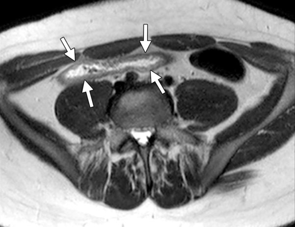
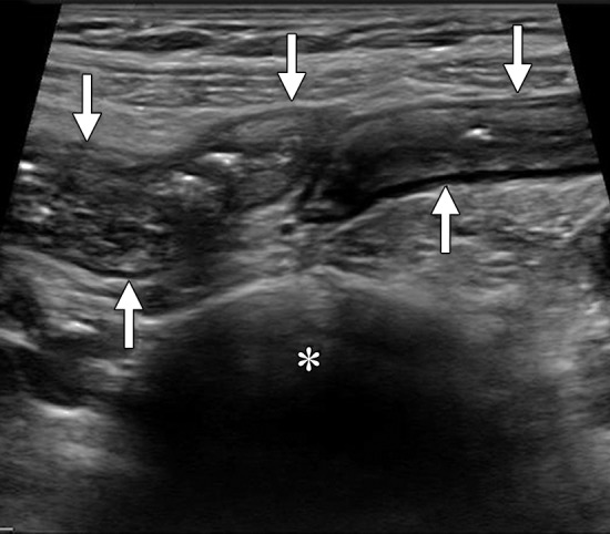
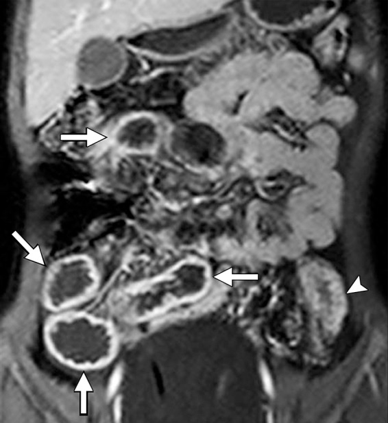
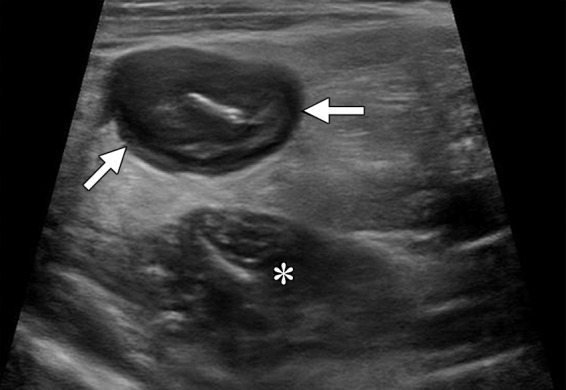
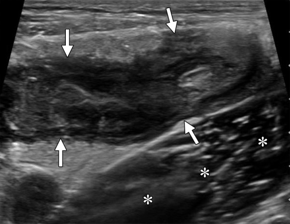
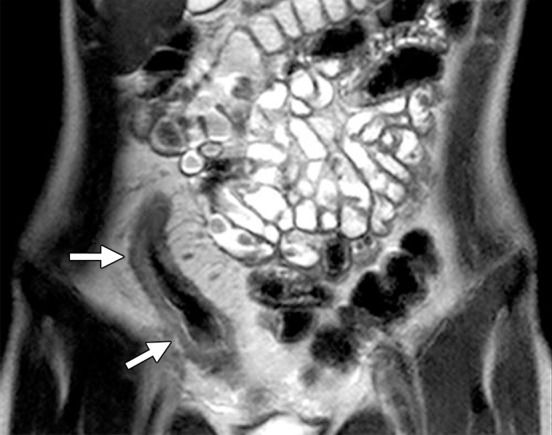
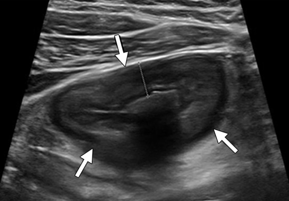
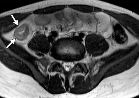
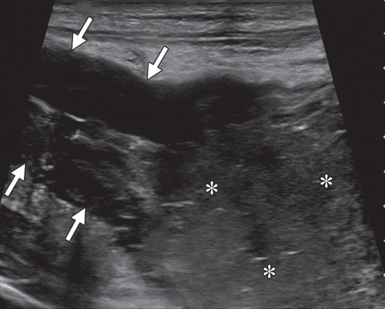
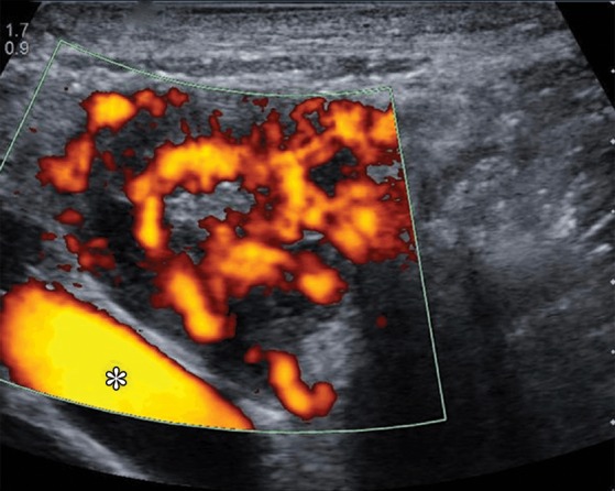
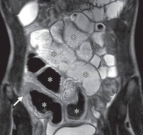
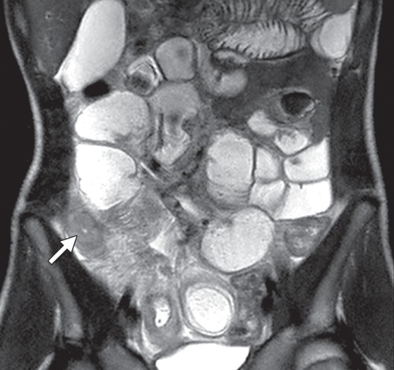
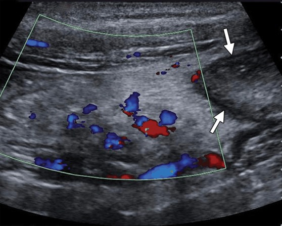
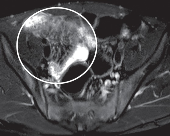
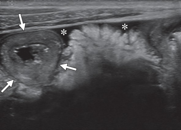
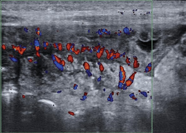
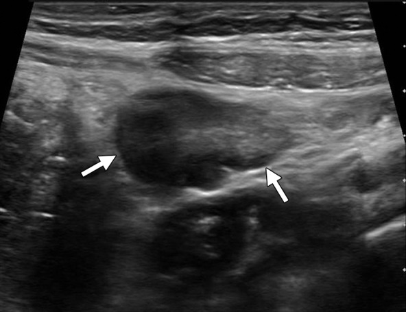
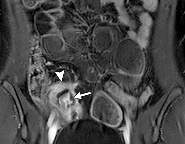
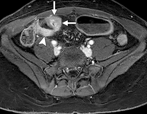
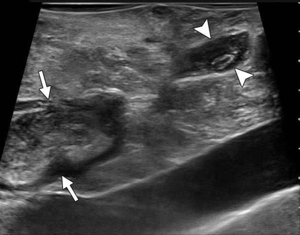
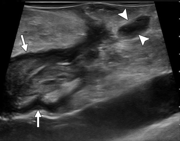
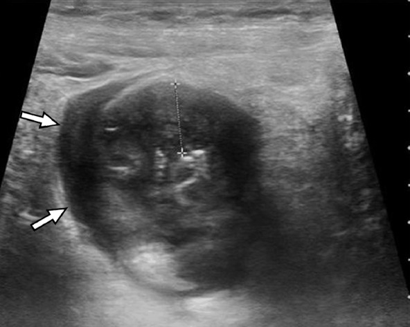
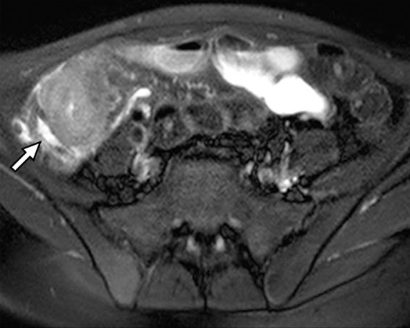
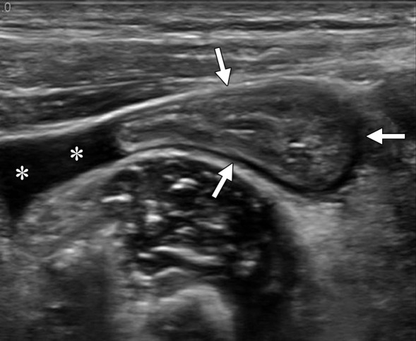
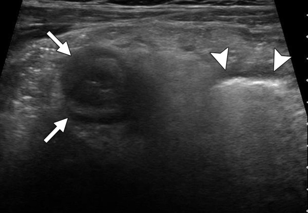
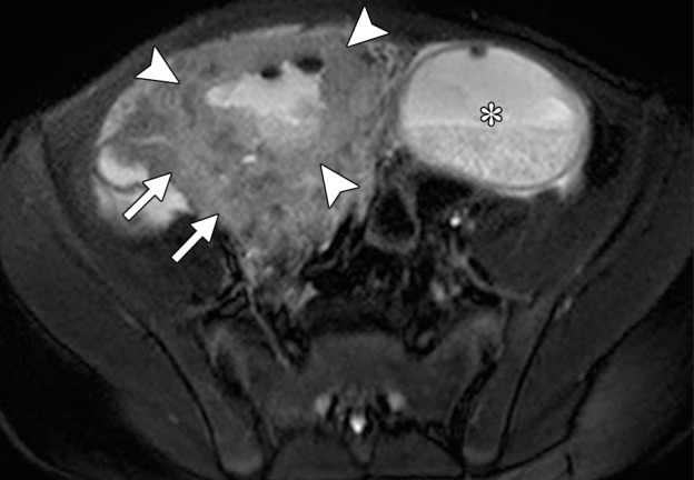
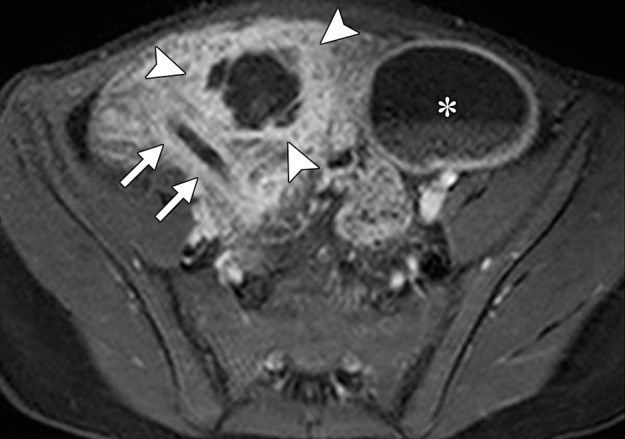
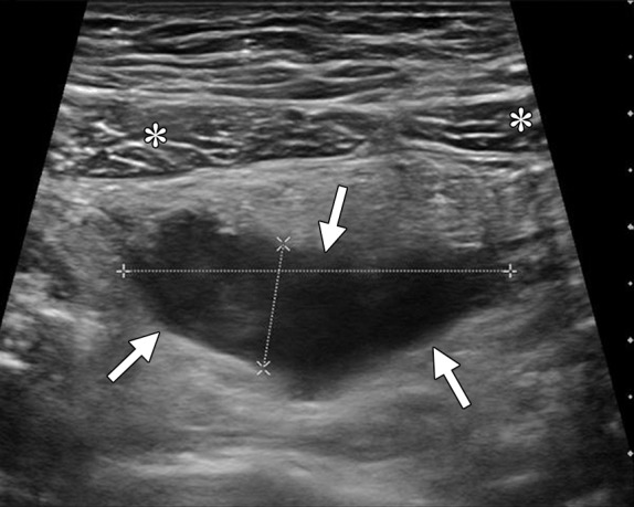
Small bowel Crohn disease is commonly diagnosed during the pediatric period, and recent investigations show that its incidence is increasing in this age group. Diagnosis and follow-up of this condition are commonly based on a combination of patient history and physical examination, disease activity surveys, laboratory assessment, and endoscopy with biopsy, but imaging also plays a central role. Ultrasonography (US) is an underutilized well-tolerated imaging modality for screening and follow-up of small bowel Crohn disease in children and adolescents. US has numerous advantages over computed tomographic (CT) enterography and magnetic resonance (MR) enterography, including low cost and no required use of oral or intravenous contrast material. US also has the potential to provide images with higher spatial resolution than those obtained at CT enterography and MR enterography, allows faster examination than does MR enterography, does not involve ionizing radiation, and does not require sedation or general anesthesia. US accurately depicts small bowel and mesenteric changes related to pediatric Crohn disease, and US findings show a high correlation with MR imaging findings in this patient population.


Figures

Figure 1a
Figure 1a
Normal terminal ileum in a 10-year-old boy. Long-axis gray-scale (a) and color Doppler (b) US images show the normal thin-walled terminal ileum (arrows) draped over the right iliac vessels (*). The lack of demonstrable blood flow seen in the terminal ileum in b is a normal finding. C = cecum.
Figure 1b
Figure 1b
Normal terminal ileum in a 10-year-old boy. Long-axis gray-scale (a) and color Doppler (b) US images show the normal thin-walled terminal ileum (arrows) draped over the right iliac vessels (*). The lack of demonstrable blood flow seen in the terminal ileum in b is a normal finding. C = cecum.
Figure 2
Figure 2
Long-axis gray-scale ex vivo US image of a resected terminal ileal specimen from an adolescent with Crohn disease. Imaging was performed with the bowel resting on a standoff pad (*). The bowel wall is thickened, has a preserved stratified appearance, and is surrounded by the thickened echogenic mesentery (creeping fat). Bowel wall layers seen are the echogenic mucosa-lumen interface (1); the hypoechoic mucosa, including the muscularis mucosae (2); the echogenic submucosa (3); and the hypoechoic muscularis propria (4).
Figure 3a
Figure 3a
Terminal ileitis due to newly diagnosed Crohn disease in a 15-year-old male adolescent. (a) Long-axis gray-scale US image shows the thick-walled stratified terminal ileum (arrows) overlying the iliac vessels (*). The echogenic submucosa is the thickest layer of the bowel wall. The abnormal bowel segment can be seen despite the patient’s large body habitus. (b) Axial single-shot fast spin-echo (FSE) MR image shows similar findings. The distal ileum (arrows) is seen immediately between the anterior abdominal wall and the right common iliac vessels.
Figure 3b
Figure 3b
Terminal ileitis due to newly diagnosed Crohn disease in a 15-year-old male adolescent. (a) Long-axis gray-scale US image shows the thick-walled stratified terminal ileum (arrows) overlying the iliac vessels (*). The echogenic submucosa is the thickest layer of the bowel wall. The abnormal bowel segment can be seen despite the patient’s large body habitus. (b) Axial single-shot fast spin-echo (FSE) MR image shows similar findings. The distal ileum (arrows) is seen immediately between the anterior abdominal wall and the right common iliac vessels.
Figure 4a
Figure 4a
Crohn disease involving the small and large bowel in a 6-year-old girl. (a) Long-axis gray-scale US image shows thickened small bowel loops (arrows) adjacent to the urinary bladder (*) with partial loss of mural layering. (b) Coronal contrast-enhanced fat-saturated T1-weighted three-dimensional (3D) MR image confirms long-segment active terminal ileitis with wall thickening, hyperenhancement, and vasa recta engorgement (arrows). The sigmoid colon in the left lower quadrant (arrowhead) is also inflamed.
Figure 4b
Figure 4b
Crohn disease involving the small and large bowel in a 6-year-old girl. (a) Long-axis gray-scale US image shows thickened small bowel loops (arrows) adjacent to the urinary bladder (*) with partial loss of mural layering. (b) Coronal contrast-enhanced fat-saturated T1-weighted three-dimensional (3D) MR image confirms long-segment active terminal ileitis with wall thickening, hyperenhancement, and vasa recta engorgement (arrows). The sigmoid colon in the left lower quadrant (arrowhead) is also inflamed.
Figure 5a
Figure 5a
Terminal ileitis due to newly diagnosed Crohn disease in a 10-year-old boy. (a, b) Short-axis (a) and long-axis (b) gray-scale US images of a bowel loop overlying the right psoas muscle (*) show marked wall thickening with loss of mural layering (arrows). (c) Coronal single-shot FSE MR image confirms distal and ileal wall thickening (arrows), with adjacent mesenteric fibrofatty proliferation and vasa recta engorgement.
Figure 5b
Figure 5b
Terminal ileitis due to newly diagnosed Crohn disease in a 10-year-old boy. (a, b) Short-axis (a) and long-axis (b) gray-scale US images of a bowel loop overlying the right psoas muscle (*) show marked wall thickening with loss of mural layering (arrows). (c) Coronal single-shot FSE MR image confirms distal and ileal wall thickening (arrows), with adjacent mesenteric fibrofatty proliferation and vasa recta engorgement.
Figure 5c
Figure 5c
Terminal ileitis due to newly diagnosed Crohn disease in a 10-year-old boy. (a, b) Short-axis (a) and long-axis (b) gray-scale US images of a bowel loop overlying the right psoas muscle (*) show marked wall thickening with loss of mural layering (arrows). (c) Coronal single-shot FSE MR image confirms distal and ileal wall thickening (arrows), with adjacent mesenteric fibrofatty proliferation and vasa recta engorgement.
Figure 6a
Figure 6a
Terminal ileitis in a 17-year-old girl with known Crohn disease. (a) Short-axis gray-scale US image through the right lower quadrant shows marked terminal ileal wall thickening (arrows), with a maximum wall thickness of 8 mm. (b) Axial single-shot FSE MR image confirms terminal ileal wall thickening (arrows).
Figure 6b
Figure 6b
Terminal ileitis in a 17-year-old girl with known Crohn disease. (a) Short-axis gray-scale US image through the right lower quadrant shows marked terminal ileal wall thickening (arrows), with a maximum wall thickness of 8 mm. (b) Axial single-shot FSE MR image confirms terminal ileal wall thickening (arrows).
Figure 7a
Figure 7a
Terminal ileitis in a 10-year-old boy with newly diagnosed Crohn disease. (a) Color Doppler US image shows terminal ileal wall thickening, with markedly increased intramural signal intensity due to active inflammation. Extensive signal aliasing is also seen. (b) Axial diffusion-weighted MR image confirms marked bowel wall inflammation (arrows). The hyperintense appearance of the bowel wall is due to restricted diffusion of water.
Figure 7b
Figure 7b
Terminal ileitis in a 10-year-old boy with newly diagnosed Crohn disease. (a) Color Doppler US image shows terminal ileal wall thickening, with markedly increased intramural signal intensity due to active inflammation. Extensive signal aliasing is also seen. (b) Axial diffusion-weighted MR image confirms marked bowel wall inflammation (arrows). The hyperintense appearance of the bowel wall is due to restricted diffusion of water.
Figure 8a
Figure 8a
Terminal ileitis in a 14-year-old girl. (a) Color Doppler US image shows mild terminal ileal wall thickening and marked intramural hyperemia due to active inflammation. The terminal ileum overlies the right iliopsoas muscle (arrow) and iliac vessels (*). (b) Axial fat-saturated single-shot FSE MR image shows 15 cm of mildly thickened distal and terminal ileum (circled), a finding that avidly enhanced at contrast-enhanced MR imaging (not shown). Arrow = right iliopsoas muscle, * = iliac vessels.
Figure 8b
Figure 8b
Terminal ileitis in a 14-year-old girl. (a) Color Doppler US image shows mild terminal ileal wall thickening and marked intramural hyperemia due to active inflammation. The terminal ileum overlies the right iliopsoas muscle (arrow) and iliac vessels (*). (b) Axial fat-saturated single-shot FSE MR image shows 15 cm of mildly thickened distal and terminal ileum (circled), a finding that avidly enhanced at contrast-enhanced MR imaging (not shown). Arrow = right iliopsoas muscle, * = iliac vessels.
Figure 9a
Figure 9a
Stricture in a 15-year-old boy with newly diagnosed Crohn disease. (a) Long-axis gray-scale US image shows an obstructing stricture. The terminal ileum is thick walled (arrows) with luminal narrowing, while the more proximal small bowel is dilated and contains debris (*). (b) Short-axis power Doppler US image shows the narrowed bowel segment with marked hyperemia due to active inflammation. * = iliac artery. (c, d) Coronal single-shot FSE MR images obtained at different levels confirm small bowel obstruction, with a thickened narrowed terminal ileum (arrow) and proximal luminal dilatation (* in c).
Figure 9b
Figure 9b
Stricture in a 15-year-old boy with newly diagnosed Crohn disease. (a) Long-axis gray-scale US image shows an obstructing stricture. The terminal ileum is thick walled (arrows) with luminal narrowing, while the more proximal small bowel is dilated and contains debris (*). (b) Short-axis power Doppler US image shows the narrowed bowel segment with marked hyperemia due to active inflammation. * = iliac artery. (c, d) Coronal single-shot FSE MR images obtained at different levels confirm small bowel obstruction, with a thickened narrowed terminal ileum (arrow) and proximal luminal dilatation (* in c).
Figure 9c
Figure 9c
Stricture in a 15-year-old boy with newly diagnosed Crohn disease. (a) Long-axis gray-scale US image shows an obstructing stricture. The terminal ileum is thick walled (arrows) with luminal narrowing, while the more proximal small bowel is dilated and contains debris (*). (b) Short-axis power Doppler US image shows the narrowed bowel segment with marked hyperemia due to active inflammation. * = iliac artery. (c, d) Coronal single-shot FSE MR images obtained at different levels confirm small bowel obstruction, with a thickened narrowed terminal ileum (arrow) and proximal luminal dilatation (* in c).
Figure 9d
Figure 9d
Stricture in a 15-year-old boy with newly diagnosed Crohn disease. (a) Long-axis gray-scale US image shows an obstructing stricture. The terminal ileum is thick walled (arrows) with luminal narrowing, while the more proximal small bowel is dilated and contains debris (*). (b) Short-axis power Doppler US image shows the narrowed bowel segment with marked hyperemia due to active inflammation. * = iliac artery. (c, d) Coronal single-shot FSE MR images obtained at different levels confirm small bowel obstruction, with a thickened narrowed terminal ileum (arrow) and proximal luminal dilatation (* in c).
Figure 10a
Figure 10a
Active terminal ileitis in a 14-year-old boy. (a) Color Doppler US image shows a thickened loop of small bowel (arrows) in the right lower quadrant, with adjacent mesenteric hyperemia and fibrofatty proliferation. (b) Axial fat-saturated single-shot FSE MR image shows numerous engorged vessels (circled) in the right lower quadrant of the small bowel mesentery adjacent to the terminal ileum.
Figure 10b
Figure 10b
Active terminal ileitis in a 14-year-old boy. (a) Color Doppler US image shows a thickened loop of small bowel (arrows) in the right lower quadrant, with adjacent mesenteric hyperemia and fibrofatty proliferation. (b) Axial fat-saturated single-shot FSE MR image shows numerous engorged vessels (circled) in the right lower quadrant of the small bowel mesentery adjacent to the terminal ileum.
Figure 11a
Figure 11a
Active terminal ileitis in a 5-year-old boy. (a) Short-axis gray-scale US image shows small bowel wall thickening (arrows) in the right lower quadrant and adjacent free fluid (*) that is likely reactive. (b) Long-axis color Doppler US image shows marked bowel wall and mesenteric hyperemia and the “comb” sign.
Figure 11b
Figure 11b
Active terminal ileitis in a 5-year-old boy. (a) Short-axis gray-scale US image shows small bowel wall thickening (arrows) in the right lower quadrant and adjacent free fluid (*) that is likely reactive. (b) Long-axis color Doppler US image shows marked bowel wall and mesenteric hyperemia and the “comb” sign.
Figure 12a
Figure 12a
Terminal ileitis in a 10-year-old boy with newly diagnosed Crohn disease. (a) Short-axis gray-scale US image shows the thick-walled heterogeneous terminal ileum in the right lower quadrant. The adjacent small bowel mesentery is markedly thickened and echogenic, a finding due to fibrofatty proliferation (*). The adjacent bowel loops are displaced. (b) Axial single-shot FSE MR image shows similar findings. The increased signal intensity surrounding the terminal ileum is due to creeping fat (*).
Figure 12b
Figure 12b
Terminal ileitis in a 10-year-old boy with newly diagnosed Crohn disease. (a) Short-axis gray-scale US image shows the thick-walled heterogeneous terminal ileum in the right lower quadrant. The adjacent small bowel mesentery is markedly thickened and echogenic, a finding due to fibrofatty proliferation (*). The adjacent bowel loops are displaced. (b) Axial single-shot FSE MR image shows similar findings. The increased signal intensity surrounding the terminal ileum is due to creeping fat (*).
Figure 13a
Figure 13a
Small bowel Crohn disease in a 10-year-old boy. (a) Gray-scale US image shows a mildly enlarged, 9-mm, likely reactive lymph node (arrows) in the small bowel mesentery. (b) Axial fat-saturated single-shot FSE MR image shows extensive signal intensity abnormality in the right lower quadrant (circled) due to inflammation, which obscures typically hyperintense lymph nodes.
Figure 13b
Figure 13b
Small bowel Crohn disease in a 10-year-old boy. (a) Gray-scale US image shows a mildly enlarged, 9-mm, likely reactive lymph node (arrows) in the small bowel mesentery. (b) Axial fat-saturated single-shot FSE MR image shows extensive signal intensity abnormality in the right lower quadrant (circled) due to inflammation, which obscures typically hyperintense lymph nodes.
Figure 14a
Figure 14a
Crohn disease in a 7-year-old boy. (a) Gray-scale US image shows an enlarged, likely reactive lymph node (arrows) in the small bowel mesentery just anterior to the right psoas muscle. The fatty hilum of the lymph node is preserved. (b) Axial diffusion-weighted MR image depicts the same lymph node (arrow).
Figure 14b
Figure 14b
Crohn disease in a 7-year-old boy. (a) Gray-scale US image shows an enlarged, likely reactive lymph node (arrows) in the small bowel mesentery just anterior to the right psoas muscle. The fatty hilum of the lymph node is preserved. (b) Axial diffusion-weighted MR image depicts the same lymph node (arrow).
Figure 15a
Figure 15a
Penetrating stricturing Crohn disease in a 19-year-old woman. (a, b) Coronal (a) and axial (b) contrast-enhanced fat-saturated T1-weighted 3D MR images obtained 1 week before surgery show a fistula between the terminal ileum (arrows) and the appendix (arrowhead). (c, d) Gray-scale US images of the resected bowel specimen show a fistula tract between the thick-walled terminal ileum (arrows) and the appendix (arrowheads). Fibrofatty thickening of the mesentery is also seen.
Figure 15b
Figure 15b
Penetrating stricturing Crohn disease in a 19-year-old woman. (a, b) Coronal (a) and axial (b) contrast-enhanced fat-saturated T1-weighted 3D MR images obtained 1 week before surgery show a fistula between the terminal ileum (arrows) and the appendix (arrowhead). (c, d) Gray-scale US images of the resected bowel specimen show a fistula tract between the thick-walled terminal ileum (arrows) and the appendix (arrowheads). Fibrofatty thickening of the mesentery is also seen.
Figure 15c
Figure 15c
Penetrating stricturing Crohn disease in a 19-year-old woman. (a, b) Coronal (a) and axial (b) contrast-enhanced fat-saturated T1-weighted 3D MR images obtained 1 week before surgery show a fistula between the terminal ileum (arrows) and the appendix (arrowhead). (c, d) Gray-scale US images of the resected bowel specimen show a fistula tract between the thick-walled terminal ileum (arrows) and the appendix (arrowheads). Fibrofatty thickening of the mesentery is also seen.
Figure 15d
Figure 15d
Penetrating stricturing Crohn disease in a 19-year-old woman. (a, b) Coronal (a) and axial (b) contrast-enhanced fat-saturated T1-weighted 3D MR images obtained 1 week before surgery show a fistula between the terminal ileum (arrows) and the appendix (arrowhead). (c, d) Gray-scale US images of the resected bowel specimen show a fistula tract between the thick-walled terminal ileum (arrows) and the appendix (arrowheads). Fibrofatty thickening of the mesentery is also seen.
Figure 16a
Figure 16a
Crohn disease in an 18-year-old woman. (a) Short-axis gray-scale US image shows marked terminal ileal wall thickening and adjacent, likely reactive, free fluid (arrows). (b) Axial fat-saturated single-shot FSE MR image confirms severe terminal ileitis with bowel wall thickening, mural edema, and adjacent mesenteric inflammation. A small amount of free fluid (arrow) is seen adjacent to the terminal ileum.
Figure 16b
Figure 16b
Crohn disease in an 18-year-old woman. (a) Short-axis gray-scale US image shows marked terminal ileal wall thickening and adjacent, likely reactive, free fluid (arrows). (b) Axial fat-saturated single-shot FSE MR image confirms severe terminal ileitis with bowel wall thickening, mural edema, and adjacent mesenteric inflammation. A small amount of free fluid (arrow) is seen adjacent to the terminal ileum.
Figure 17a
Figure 17a
Crohn disease in a 4-year-old boy. (a) Short-axis gray-scale US image shows terminal ileal wall thickening (arrows) and adjacent free fluid (*). (b) Coronal single-shot FSE MR image shows wall thickening in the terminal ileum and a small amount of adjacent free fluid (arrow). The free fluid is more easily appreciated at US.
Figure 17b
Figure 17b
Crohn disease in a 4-year-old boy. (a) Short-axis gray-scale US image shows terminal ileal wall thickening (arrows) and adjacent free fluid (*). (b) Coronal single-shot FSE MR image shows wall thickening in the terminal ileum and a small amount of adjacent free fluid (arrow). The free fluid is more easily appreciated at US.
Figure 18a
Figure 18a
Terminal ileitis due to Crohn disease in a 14-year-old girl. (a) Long-axis gray-scale US image shows an ill-defined area of decreased echogenicity (arrows) between an inflamed bowel loop (arrowheads) and the anterior abdominal wall (*), a finding consistent with phlegmon. (b, c) Axial contrast-enhanced fat-saturated T1-weighted 3D MR images obtained at different levels show marked terminal ileal wall thickening and hyperenhancement (arrowheads). The ill-defined perienteric soft-tissue enhancement adjacent to the terminal ileum (arrows) is consistent with phlegmon and is likely the result of penetrating Crohn disease. No drainable fluid component was identified.
Figure 18b
Figure 18b
Terminal ileitis due to Crohn disease in a 14-year-old girl. (a) Long-axis gray-scale US image shows an ill-defined area of decreased echogenicity (arrows) between an inflamed bowel loop (arrowheads) and the anterior abdominal wall (*), a finding consistent with phlegmon. (b, c) Axial contrast-enhanced fat-saturated T1-weighted 3D MR images obtained at different levels show marked terminal ileal wall thickening and hyperenhancement (arrowheads). The ill-defined perienteric soft-tissue enhancement adjacent to the terminal ileum (arrows) is consistent with phlegmon and is likely the result of penetrating Crohn disease. No drainable fluid component was identified.
Figure 18c
Figure 18c
Terminal ileitis due to Crohn disease in a 14-year-old girl. (a) Long-axis gray-scale US image shows an ill-defined area of decreased echogenicity (arrows) between an inflamed bowel loop (arrowheads) and the anterior abdominal wall (*), a finding consistent with phlegmon. (b, c) Axial contrast-enhanced fat-saturated T1-weighted 3D MR images obtained at different levels show marked terminal ileal wall thickening and hyperenhancement (arrowheads). The ill-defined perienteric soft-tissue enhancement adjacent to the terminal ileum (arrows) is consistent with phlegmon and is likely the result of penetrating Crohn disease. No drainable fluid component was identified.
Figure 19a
Figure 19a
Abscess in an 11-year-old girl with known Crohn disease and worsening abdominal pain. (a) Gray-scale US image shows terminal ileal wall (arrows) and mesenteric thickening. A curvilinear gas-filled structure (arrowheads) in the anterior abdomen is a large abscess but could be mistaken for a bowel loop. (b, c) Axial fat-saturated single-shot FSE MR image (b) and contrast-enhanced fat-saturated T1-weighted 3D MR image (c) show a large, peripherally enhancing, gas-containing abscess (arrowheads) in the small bowel mesentery. The thick-walled narrowed terminal ileum (arrows) is difficult to visualize because of surrounding inflammation. A dilated debris-filled bowel loop (*) in the left lower quadrant is due to the obstructed small bowel.
Figure 19b
Figure 19b
Abscess in an 11-year-old girl with known Crohn disease and worsening abdominal pain. (a) Gray-scale US image shows terminal ileal wall (arrows) and mesenteric thickening. A curvilinear gas-filled structure (arrowheads) in the anterior abdomen is a large abscess but could be mistaken for a bowel loop. (b, c) Axial fat-saturated single-shot FSE MR image (b) and contrast-enhanced fat-saturated T1-weighted 3D MR image (c) show a large, peripherally enhancing, gas-containing abscess (arrowheads) in the small bowel mesentery. The thick-walled narrowed terminal ileum (arrows) is difficult to visualize because of surrounding inflammation. A dilated debris-filled bowel loop (*) in the left lower quadrant is due to the obstructed small bowel.
Figure 19c
Figure 19c
Abscess in an 11-year-old girl with known Crohn disease and worsening abdominal pain. (a) Gray-scale US image shows terminal ileal wall (arrows) and mesenteric thickening. A curvilinear gas-filled structure (arrowheads) in the anterior abdomen is a large abscess but could be mistaken for a bowel loop. (b, c) Axial fat-saturated single-shot FSE MR image (b) and contrast-enhanced fat-saturated T1-weighted 3D MR image (c) show a large, peripherally enhancing, gas-containing abscess (arrowheads) in the small bowel mesentery. The thick-walled narrowed terminal ileum (arrows) is difficult to visualize because of surrounding inflammation. A dilated debris-filled bowel loop (*) in the left lower quadrant is due to the obstructed small bowel.
Figure 20a
Figure 20a
Abscess in a 16-year-old girl with newly diagnosed Crohn disease. (a) Transverse gray-scale US image shows a 3-cm hypoechoic fluid collection (arrows) in the greater omentum, located just deep to the abdominal wall musculature (*). (b, c) Axial (b) and coronal (c) contrast-enhanced fat-saturated T1-weighted 3D MR images show extensive inflammation of the greater omentum and small bowel mesentery. A peripherally enhancing round structure (arrow) located just deep to the abdominal wall musculature (* in b) corresponds to the abnormality seen in a. This small focal abscess is better appreciated at US than at MR imaging.
Figure 20b
Figure 20b
Abscess in a 16-year-old girl with newly diagnosed Crohn disease. (a) Transverse gray-scale US image shows a 3-cm hypoechoic fluid collection (arrows) in the greater omentum, located just deep to the abdominal wall musculature (*). (b, c) Axial (b) and coronal (c) contrast-enhanced fat-saturated T1-weighted 3D MR images show extensive inflammation of the greater omentum and small bowel mesentery. A peripherally enhancing round structure (arrow) located just deep to the abdominal wall musculature (* in b) corresponds to the abnormality seen in a. This small focal abscess is better appreciated at US than at MR imaging.
Figure 20c
Figure 20c
Abscess in a 16-year-old girl with newly diagnosed Crohn disease. (a) Transverse gray-scale US image shows a 3-cm hypoechoic fluid collection (arrows) in the greater omentum, located just deep to the abdominal wall musculature (*). (b, c) Axial (b) and coronal (c) contrast-enhanced fat-saturated T1-weighted 3D MR images show extensive inflammation of the greater omentum and small bowel mesentery. A peripherally enhancing round structure (arrow) located just deep to the abdominal wall musculature (* in b) corresponds to the abnormality seen in a. This small focal abscess is better appreciated at US than at MR imaging.

Similar articles

Cited by

References

    1. Day AS, Ledder O, Leach ST, Lemberg DA. Crohn’s and colitis in children and adolescents. World J Gastroenterol 2012;18(41):5862–5869. - PMC - PubMed
    1. Rabizadeh S, Dubinsky M. Update in pediatric inflammatory bowel disease. Rheum Dis Clin North Am 2013;39(4): 789–799. - PMC - PubMed
    1. Yu AP, Cabanilla LA, Wu EQ, Mulani PM, Chao J. The costs of Crohn’s disease in the United States and other Western countries: a systematic review. Curr Med Res Opin 2008;24(2):319–328. - PubMed
    1. Benitez JM, Meuwis MA, Reenaers C, Van Kemseke C, Meunier P, Louis E. Role of endoscopy, cross-sectional imaging and biomarkers in Crohn’s disease monitoring. Gut 2013;62(12):1806–1816. - PubMed
    1. Domina JG, Dillman JR, Adler J, et al. . Imaging trends and radiation exposure in pediatric inflammatory bowel disease at an academic children’s hospital. AJR Am J Roentgenol 2013;201(1):W133–W140. - PubMed

Publication types

Supplementary concepts

LinkOut - more resources