. 2015 Apr 28;112(9):1536-45.
doi: 10.1038/bjc.2015.113. Epub 2015 Apr 2.

Low-temperature plasma treatment induces DNA damage leading to necrotic cell death in primary prostate epithelial cells

Affiliations

Low-temperature plasma treatment induces DNA damage leading to necrotic cell death in primary prostate epithelial cells

A M Hirst et al. Br J Cancer. .

Abstract

Background: In recent years, the rapidly advancing field of low-temperature atmospheric pressure plasmas has shown considerable promise for future translational biomedical applications, including cancer therapy, through the generation of reactive oxygen and nitrogen species.

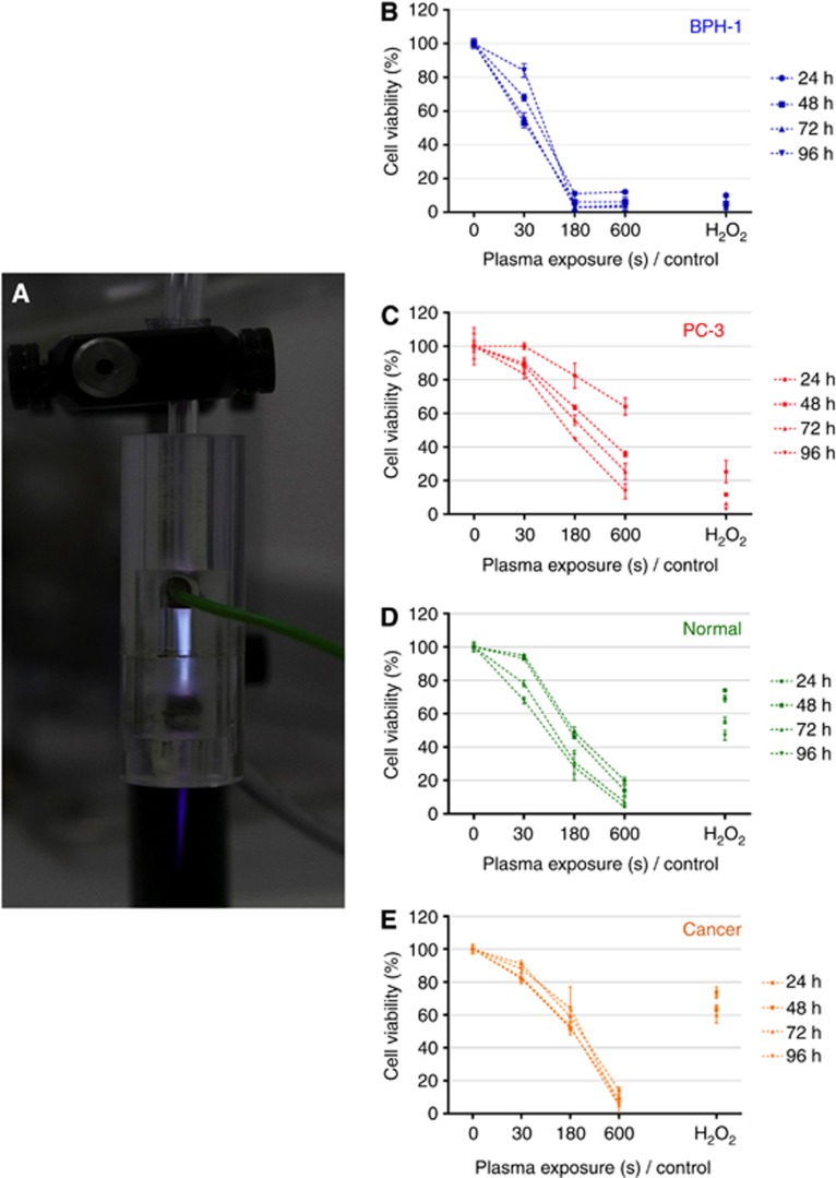
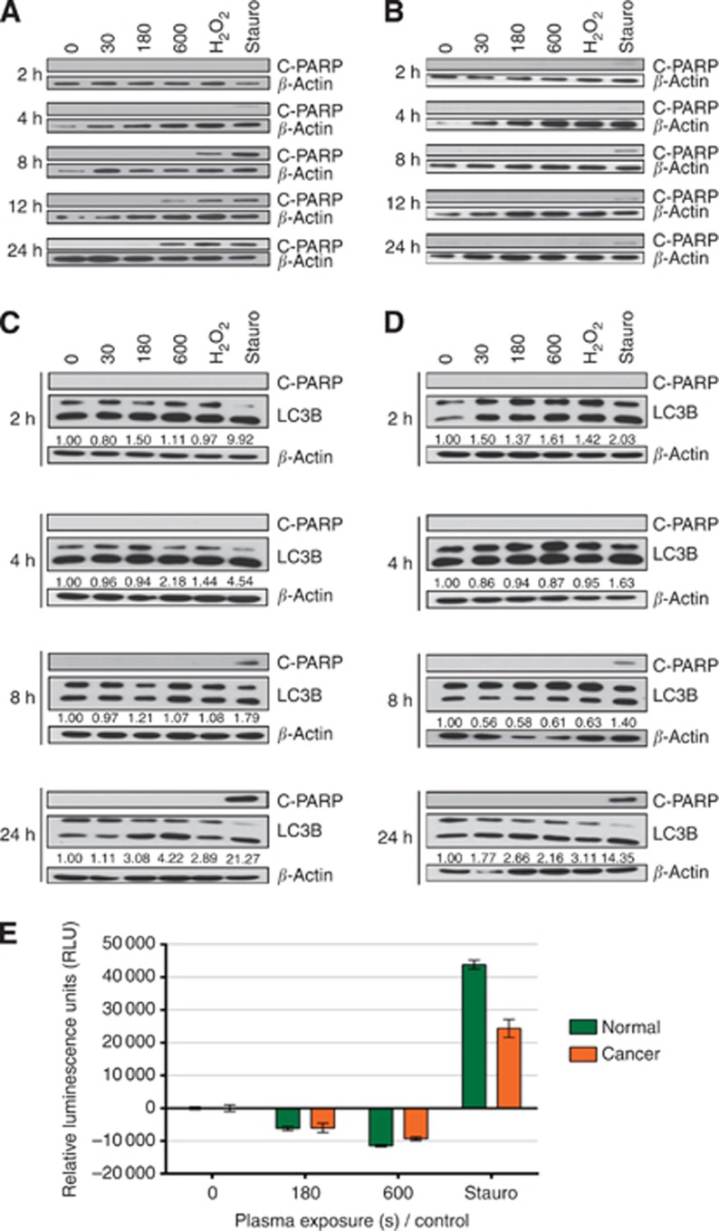
Method: The cytopathic effect of low-temperature plasma was first verified in two commonly used prostate cell lines: BPH-1 and PC-3 cells. The study was then extended to analyse the effects in paired normal and tumour (Gleason grade 7) prostate epithelial cells cultured directly from patient tissue. Hydrogen peroxide (H2O2) and staurosporine were used as controls throughout.

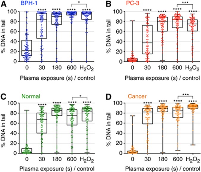
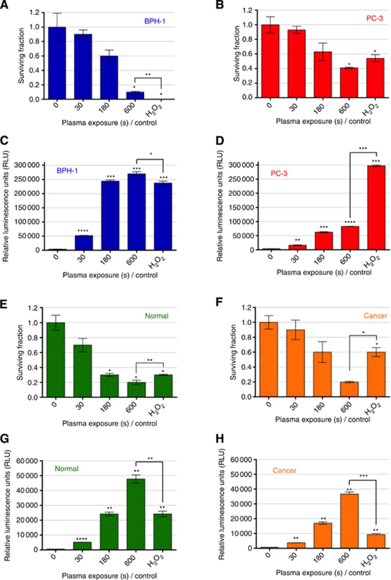
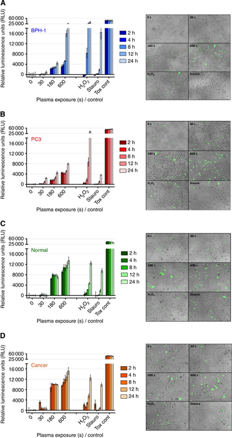
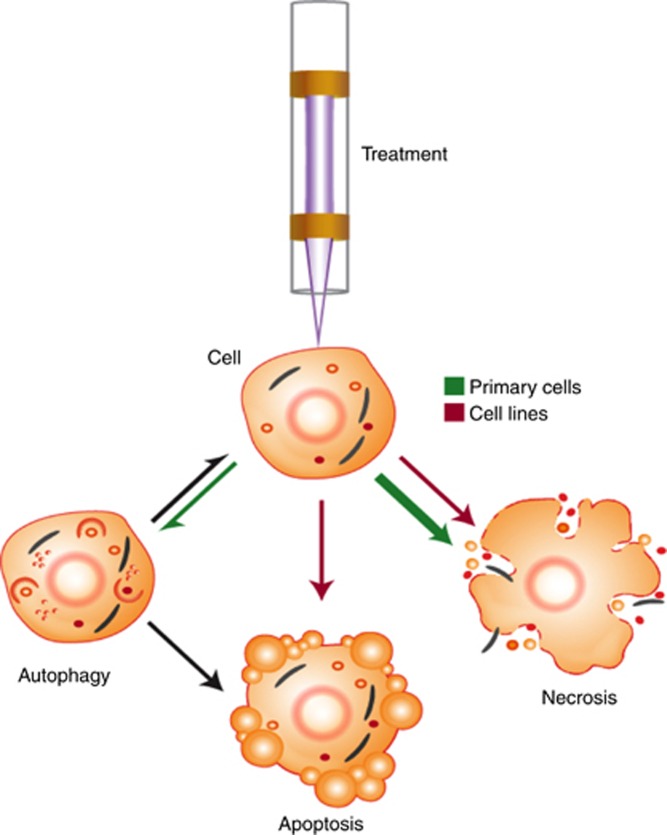
Results: Low-temperature plasma (LTP) exposure resulted in high levels of DNA damage, a reduction in cell viability, and colony-forming ability. H2O2 formed in the culture medium was a likely facilitator of these effects. Necrosis and autophagy were recorded in primary cells, whereas cell lines exhibited apoptosis and necrosis.

Conclusions: This study demonstrates that LTP treatment causes cytotoxic insult in primary prostate cells, leading to rapid necrotic cell death. It also highlights the need to study primary cultures in order to gain more realistic insight into patient response.


Figures

Figure 1
Figure 1
LTP treatment leads to a reduction in cell viability. (A) Cells were treated with a dielectric barrier discharge LTP jet, using 2 SLM helium flow rate with 0.3% O2 admixture, operated at 6 kV and 30 kHz. Reduction in cell viability was determined by alamarBlue assay (B, C) in BPH-1 and PC-3 cells and (D, E) in normal and cancer primary epithelial cells at 24, 48, 72 and 96 h postexposure.
Figure 2
Figure 2
LTP treatment induces high levels of DNA damage in both prostate cell lines and primary epithelial cells. Cells were treated with LTP or H2O2 control (1 mM). DNA damage levels were measured using the alkaline comet assay and are represented as the percentage of DNA-in-tail in (A) BPH-1 and (B) PC-3 cells and in (C) normal and (D) cancer primary epithelial cells. Each dot represents a single cell, with a minimum of 100 cells counted for each exposure. Data are expressed as median±s.e. and are analysed by Mann–Whitney test. All significance was determined against untreated samples unless otherwise indicated.
Figure 3
Figure 3
LTP treatment leads to reduction in colony-forming efficiency, and high levels of H2O2 in cell culture media. Cells were treated with LTP or H2O2 control (1 mM). Cell recovery was quantified using the colony-forming assays and is represented as surviving fraction posttreatment in (A) BPH-1 and (B) PC-3 cells and in (E) normal and (F) cancer primary epithelial cells. Immediately following treatment, ROS-Glo H2O2 luminescence assay (Promega) was performed to ascertain H2O2 levels in the culture media of (C) BPH-1 and (D) PC-3 cells and in (G) normal and (H) cancer primary epithelial cells (Note the different y axis scales). Data are expressed as mean±s.e., with statistical analysis conducted using unpaired t-test with Welch's correction. All significance was determined against untreated samples unless otherwise indicated.
Figure 4
Figure 4
LTP treatment leads to necrotic cell death in both prostate cell lines and primary epithelial cells. Immediately following LTP treatment, the CellTox green cytoxicity assay (Promega) was performed to identify cells with comprimised membrane integrity characteristic of necrosis. In all, 1 mM H2O2, 1 μM staurosporine, and cell lysing agent (supplied with assay) were used as controls. Fluorescence intensity was quantified at 2, 4, 8, 12, and 24 h following treatment and normalised to untreated control wells in (A) BPH-1 and (B) PC-3 cell lines and in (C) normal and (D) cancer primary epithelial cells. Data are expressed as mean±s.e. Supporting fluorescence microscopy images ( × 10 magnification) taken at 4 h after treatment are also shown for each cell type.
Figure 5
Figure 5
Cell death mechanisms following LTP treatment vary between cell types. Following treatment with LTP, 1 mM H2O2, or 1 μM staurosporine, protein lysates were harvested. (A) BPH-1 and (B) PC-3 cell line lysates were probed for apoptosis (C-PARP) by western blotting. (C) Normal and (D) cancer primary epithelial lysates were probed for apoptosis (C-PARP) and also autophagy (LC3B I/II). β-Actin was used as a loading control throughout. Band intensity quantification was performed using the ImageJ software. Further analysis of apoptotic activity was conducted in (E) primary epithelial cells using Caspase-glo 3/7 assay (Promega). Immediately following treatment, caspase-glo 3/7 detection reagent was added to all wells, and luminescence intensity was quantified at 24 h. Readings were normalised to untreated control and are expressed as mean±s.e.
Figure 6
Figure 6
Overview of cellular response mechanism following LTP treatment. As a result of exposure to LTP, cells were observed to undergo (or a combination of) autophagy, apoptosis or necrosis. The relative proportions of, and differences between, cell lines (red arrows) and primary epithelial cells (green arrows) that exhibit these phenomena is emphasised. Adapted from Kepp et al (2011).

References

    1. Ahn HJ, Kim KI, Hoan NN, Kim CH, Moon E, Choi KS, Yang SS, Lee JS. Targeting cancer cells with reactive oxygen and nitrogen species generated by atmospheric-pressure air plasma. PLoS One. 2014;9:e86173. - PMC - PubMed
    1. Bandyopadhyay U, Das D, Banerjee RK. Reactive oxygen species: oxidative damage and pathogenesis. Curr Sci. 1999;77:658–666.
    1. Casiano CA, Ochs RL, Tan EM. Distinct cleavage products of nuclear proteins in apoptosis and necrosis revealed by autoantibody probes. Cell Death Differ. 1998;5:183–190. - PubMed
    1. Chang JW, Kang SU, Shin YS, Kim KI, Seo SJ, Yang SS, Lee JS, Moon E, Baek SJ, Lee K, Kim CH. Non-thermal atmospheric pressure plasma induces apoptosis in oral cavity squamous cell carcinoma: Involvement of DNA-damage-triggering sub-G(1) arrest via the ATM/p53 pathway. Arch Biochem Biophys. 2014;545:133–140. - PubMed
    1. Chen MJ, Weltman E, Hanriot RM, Luz FP, Cecilio PJ, Da Cruz JC, Moreira FR, Santos AS, Martins LC, Nadalin W. Intensity modulated radiotherapy for localized prostate cancer: rigid compliance to dose-volume constraints as a warranty of acceptable toxicity. Radiat Oncol. 2007;2:6. - PMC - PubMed

Publication types