

Plastid genome-based phylogeny pinpointed the origin of the green-colored plastid in the dinoflagellate Lepidodinium chlorophorum
- PMID: 25840416
- PMCID: PMC4419806
- DOI: 10.1093/gbe/evv060
Plastid genome-based phylogeny pinpointed the origin of the green-colored plastid in the dinoflagellate Lepidodinium chlorophorum
Abstract
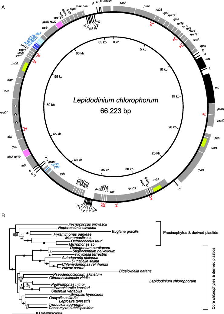
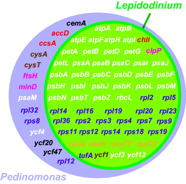
Unlike many other photosynthetic dinoflagellates, whose plastids contain a characteristic carotenoid peridinin, members of the genus Lepidodinium are the only known dinoflagellate species possessing green alga-derived plastids. However, the precise origin of Lepidodinium plastids has hitherto remained uncertain. In this study, we completely sequenced the plastid genome of Lepidodinium chlorophorum NIES-1868. Our phylogenetic analyses of 52 plastid-encoded proteins unite L. chlorophorum exclusively with a pedinophyte, Pedinomonas minor, indicating that the green-colored plastids in Lepidodinium spp. were derived from an endosymbiotic pedinophyte or a green alga closely related to pedinophytes. Our genome comparison incorporating the origin of the Lepidodinium plastids strongly suggests that the endosymbiont plastid genome acquired by the ancestral Lepidodinium species has lost genes encoding proteins involved in metabolism and biosynthesis, protein/metabolite transport, and plastid division during the endosymbiosis. We further discuss the commonalities and idiosyncrasies in genome evolution between the L. chlorophorum plastid and other plastids acquired through endosymbiosis of eukaryotic photoautotrophs.
Keywords: Lepidodinium; Pedinophyceae; genome reduction; plastid replacement; secondary plastids.
© The Author(s) 2015. Published by Oxford University Press on behalf of the Society for Molecular Biology and Evolution.
Figures
Similar articles
-
A phylogenetic mosaic plastid proteome and unusual plastid-targeting signals in the green-colored dinoflagellate Lepidodinium chlorophorum.BMC Evol Biol. 2010 Jun 21;10:191. doi: 10.1186/1471-2148-10-191. BMC Evol Biol. 2010. PMID: 20565933 Free PMC article.
-
Origins of plastids and glyceraldehyde-3-phosphate dehydrogenase genes in the green-colored dinoflagellate Lepidodinium chlorophorum.Gene. 2008 Feb 29;410(1):26-36. doi: 10.1016/j.gene.2007.11.008. Epub 2007 Nov 28. Gene. 2008. PMID: 18191504
-
Prasinoxanthin is absent in the green-colored dinoflagellate Lepidodinium chlorophorum strain NIES-1868: pigment composition and 18S rRNA phylogeny.J Plant Res. 2012 Nov;125(6):705-11. doi: 10.1007/s10265-012-0486-6. Epub 2012 Mar 23. J Plant Res. 2012. PMID: 22441568
-
Interpreting the complexities of the plastid genome in dinoflagellates: a mini-review of recent advances.Plant Mol Biol. 2024 Oct 21;114(6):114. doi: 10.1007/s11103-024-01511-3. Plant Mol Biol. 2024. PMID: 39432142 Review.
-
Integration of plastids with their hosts: Lessons learned from dinoflagellates.Proc Natl Acad Sci U S A. 2015 Aug 18;112(33):10247-54. doi: 10.1073/pnas.1421380112. Epub 2015 May 20. Proc Natl Acad Sci U S A. 2015. PMID: 25995366 Free PMC article. Review.
Cited by
-
Plastid phylogenomics with broad taxon sampling further elucidates the distinct evolutionary origins and timing of secondary green plastids.Sci Rep. 2018 Jan 24;8(1):1523. doi: 10.1038/s41598-017-18805-w. Sci Rep. 2018. PMID: 29367699 Free PMC article.
-
Dinoflagellates with relic endosymbiont nuclei as models for elucidating organellogenesis.Proc Natl Acad Sci U S A. 2020 Mar 10;117(10):5364-5375. doi: 10.1073/pnas.1911884117. Epub 2020 Feb 24. Proc Natl Acad Sci U S A. 2020. PMID: 32094181 Free PMC article.
-
Encyclopedia of Family A DNA Polymerases Localized in Organelles: Evolutionary Contribution of Bacteria Including the Proto-Mitochondrion.Mol Biol Evol. 2024 Feb 1;41(2):msae014. doi: 10.1093/molbev/msae014. Mol Biol Evol. 2024. PMID: 38271287 Free PMC article.
-
Patterns in evolutionary origins of heme, chlorophyll a and isopentenyl diphosphate biosynthetic pathways suggest non-photosynthetic periods prior to plastid replacements in dinoflagellates.PeerJ. 2018 Aug 3;6:e5345. doi: 10.7717/peerj.5345. eCollection 2018. PeerJ. 2018. PMID: 30083465 Free PMC article.
-
Comparative Plastid Genomics of Green-Colored Dinoflagellates Unveils Parallel Genome Compaction and RNA Editing.Front Plant Sci. 2022 Jul 11;13:918543. doi: 10.3389/fpls.2022.918543. eCollection 2022. Front Plant Sci. 2022. PMID: 35898209 Free PMC article.
References
-
- Archibald JM. The puzzle of plastid evolution. Curr Biol. 2009;19:R81–R88. - PubMed
-
- Archibald JM. The evolution of algae by secondary and tertiary endosymbiosis. Adv Bot Res. 2012;64:87–118.
-
- Boetzer M, Henkel CV, Jansen HJ, Butler D, Pirovano W. Scaffolding pre-assembled contigs using SSPACE. Bioinformatics. 2011;27:578–579. - PubMed
Publication types
MeSH terms
Associated data
- Actions
LinkOut - more resources
Full Text Sources
Other Literature Sources
Research Materials
Miscellaneous
