. 2015 Apr 2;5(6):1283-99.
doi: 10.1534/g3.114.015842.

A Multifunctional Mutagenesis System for Analysis of Gene Function in Zebrafish

Affiliations

A Multifunctional Mutagenesis System for Analysis of Gene Function in Zebrafish

Helen Ngoc Bao Quach et al. G3 (Bethesda). .

Abstract

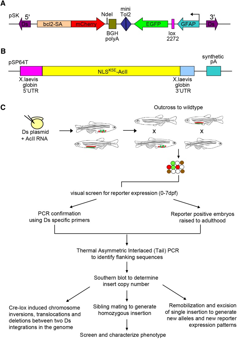
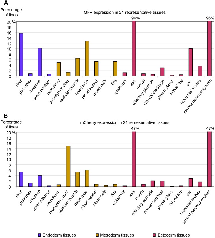
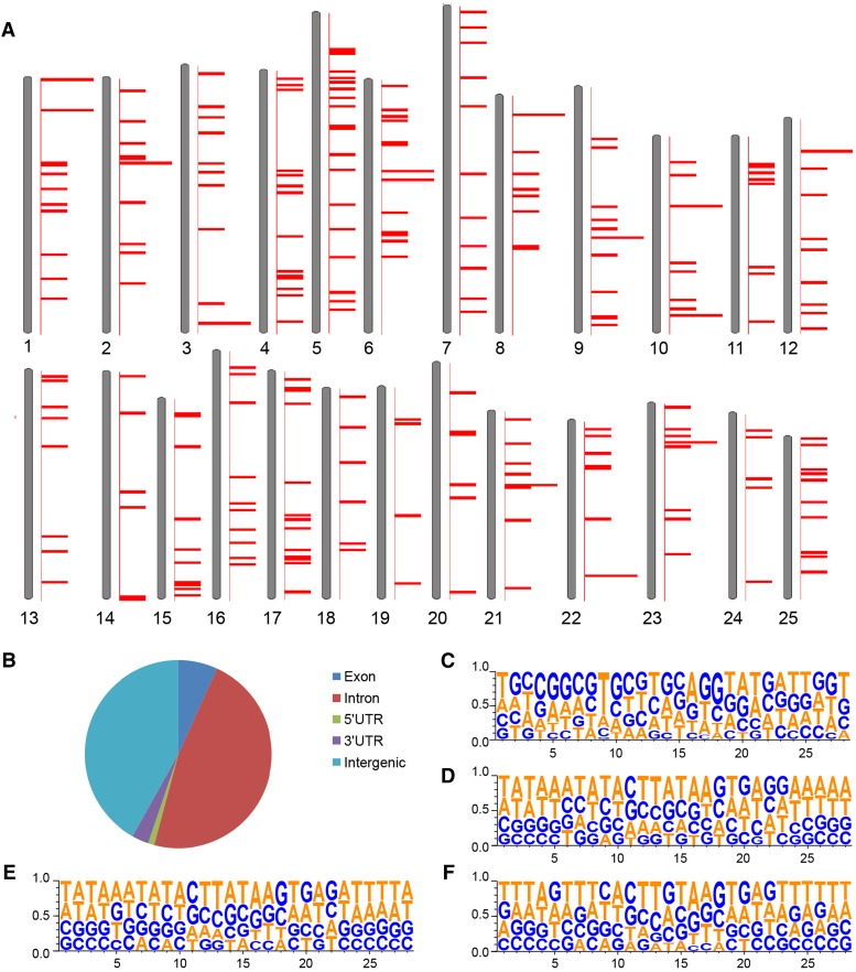
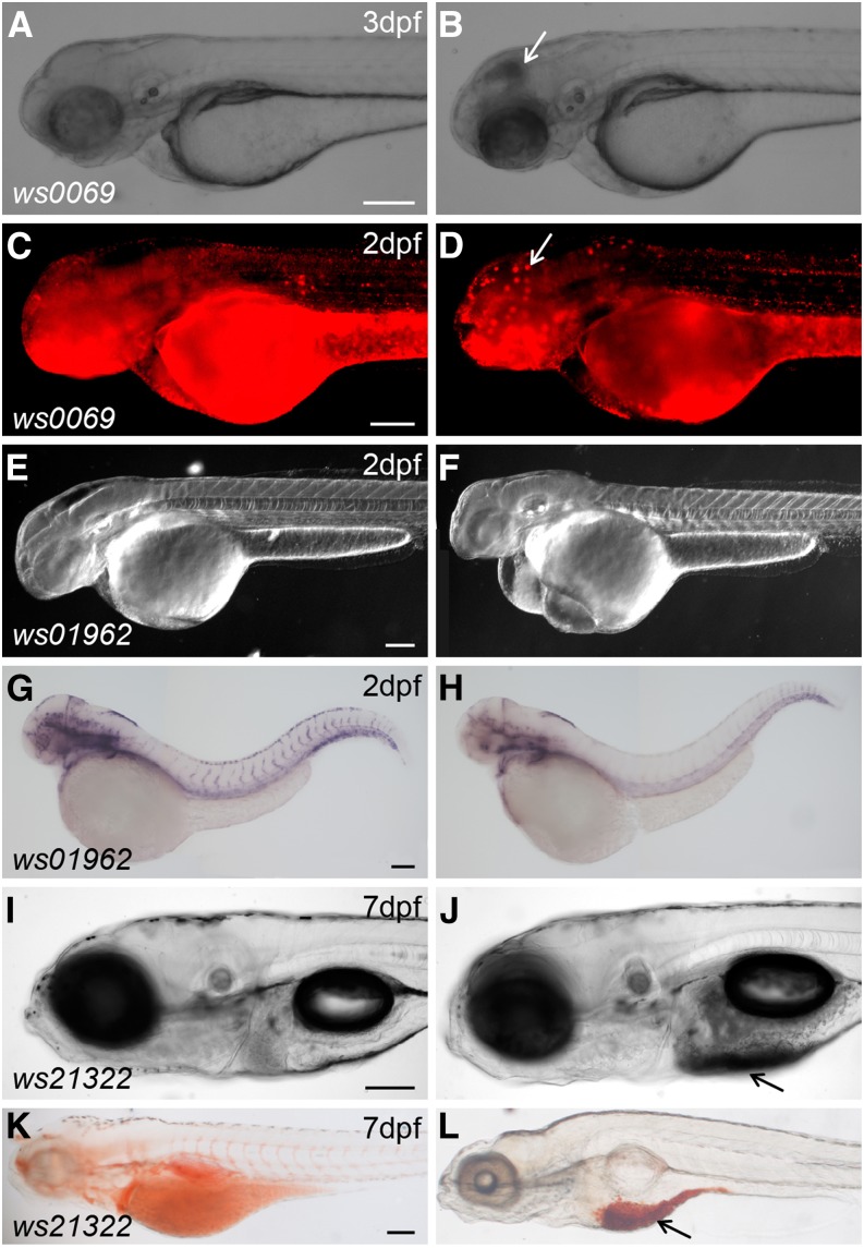
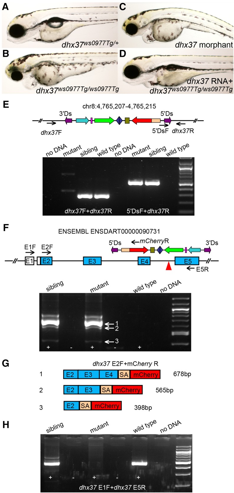
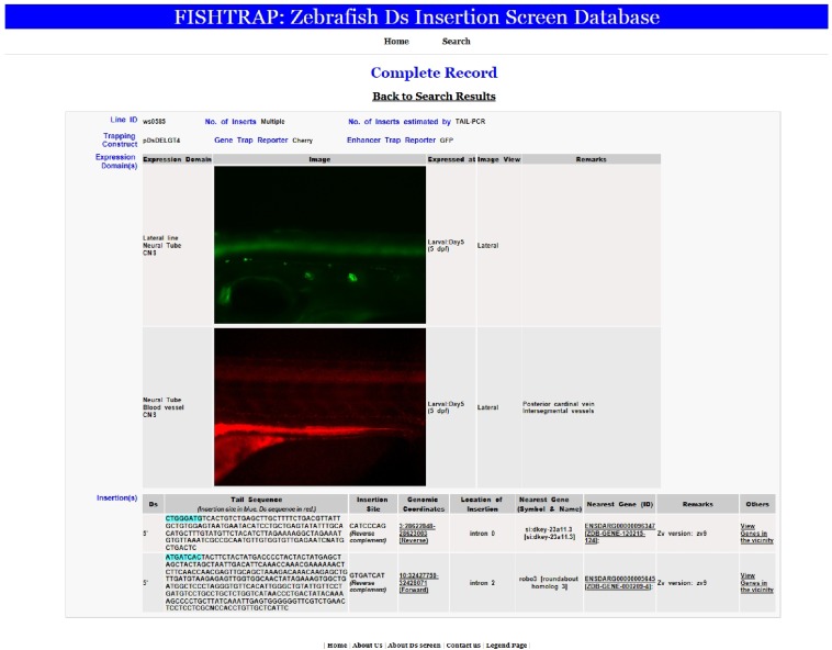
Since the sequencing of the human reference genome, many human disease-related genes have been discovered. However, understanding the functions of all the genes in the genome remains a challenge. The biological activities of these genes are usually investigated in model organisms such as mice and zebrafish. Large-scale mutagenesis screens to generate disruptive mutations are useful for identifying and understanding the activities of genes. Here, we report a multifunctional mutagenesis system in zebrafish using the maize Ds transposon. Integration of the Ds transposable element containing an mCherry reporter for protein trap events and an EGFP reporter for enhancer trap events produced a collection of transgenic lines marking distinct cell and tissue types, and mutagenized genes in the zebrafish genome by trapping and prematurely terminating endogenous protein coding sequences. We obtained 642 zebrafish lines with dynamic reporter gene expression. The characterized fish lines with specific expression patterns will be made available through the European Zebrafish Resource Center (EZRC), and a database of reporter expression is available online (http://fishtrap.warwick.ac.uk/). Our approach complements other efforts using zebrafish to facilitate functional genomic studies in this model of human development and disease.

Keywords: Ac/Ds transposon; functional genomics; gene expression; insertional mutagenesis.


Figures

Figure 1
Figure 1
Schematic representation of the multi-functional Ac/Ds transposon system and insertion screen. (A) The pDsDELGT4 vector consists of a protein trap unit and an enhancer trap unit. The protein trap unit is close to the Ds 5′ terminal repeat sequences. The mCherry coding sequence without the first methionine (red) is flanked by the zebrafish B-cell leukemia/lymphoma 2 (bcl2) splice acceptor sequence and the bovine growth hormone (BGH) polyadenylation signal. In the reverse orientation, close to the Ds 3′ terminal repeat sequences, the enhancer trap reporter GFP (green) is downstream of a short glial fibrillary acidic protein (GFAP) promoter and a lox2272 site. A mini Tol2 sequence is present between the two trap units (blue). (B) Schematic representation of the construct for synthesizing Ac transposase mRNA, with 5′UTR and 3′UTR sequences from the Xenopus globin gene. (C) Overview of the DsDELGT4 mutagenesis screen. pDsDELGT4 plasmid was co-injected with capped Ac mRNA into one-cell stage embryos (F0). Founders with transient reporter expression were raised to adulthood and mated with wild-type (AB) fish. F1 embryos were visually screened for reporter expression from fertilization until 7 d after fertilization. Ds integrations were verified by PCR using Ds specific primers. Reporter positive F1 embryos were raised to adulthood. TAIL-PCR and Southern hybridization were performed with genomic DNA isolated from the tail-fin of F1 fish and subsequent generations to map the integrations and determine the number of Ds insertions. Phenotype analysis of homozygous mutants generated by mating siblings with the same integration was performed. Cre-mediated recombination between two Ds integrations was performed to generate precise segmental deletions.
Figure 2
Figure 2
Reporter expression in different tissues. Graphs in (A) and (B) show percentage of reporter positive lines (n = 642) with enhancer trap reporter GFP expression (A) and protein trap reporter mCherry expression (B) in 21 tissues/organs derived from the three germ layers.
Figure 3
Figure 3
Reporter expression in various cell types. (A–I) Examples of enhancer trap reporter GFP expression patterns in the liver (A), pancreas (B), intestine (C), central nervous system (D), swim bladder (D), muscle cells (E), heart tube (F), branchial arches (G), fins (H), and cranial cartilage (I). (J–O) Examples of protein trap reporter mCherry expression patterns in notochord (J), anterior pronephric duct (K), mouth (L), inner ear (M), olfactory placode (N), and pineal gland (O). Ventral views are shown in (A) and (G). Lateral views are shown in (B–F), (H), (J), (K), and (M), with anterior to the left. Dorsal views are shown in (I), (L), (N), and (O). CNS, central nervous system; SB, swim bladder. Scale bars represent 100 µm in (B), 200 µm in (D), 20 µm in (J) and (M), and 50 µm in other images.
Figure 4
Figure 4
Tissue-specific expression patterns may reveal activities of trapped loci. (A–D) Bright field (A) and fluorescent images of Tg(DsDELGT4)ws2036 embryos at 52 hr after fertilization (A–C), and 5 d after fertilization (D). Arrows show GFP expression in venous sprouts from posterior cardinal vein (C) and inter-segmental veins (ISVs) (D). (E–J) Bright field (I) and fluorescent images of Tg(DsDELGT4)ws0449 embryos at 5.25 hr after fertilization (E, G), 2 d after fertilization (F, H), and 6 d after fertilization (I, J). GFP expresses in CNS and mCherry expresses in posterior cardinal vein (PCV), caudal vein (CV) (arrowheads in H), and intersegmental veins (arrows in J), but not in dorsal aorta (DA in J). (K, L) Fluorescent images of a CU928220.1ws21321Tg embryo with mCherry expression in the inner ear of a larva 7 d after fertilization (K, arrowhead) and in neuromasts of the posterior lateral line primordium (pLLP) (L, arrows) at 6 d after fertilization. Scale bars, 50 µm in (A, C, D, E, I), 250 µm in (F) and (L), and 10 µm in (K).
Figure 5
Figure 5
Mapping of DsDELGT4 integrations to zebrafish chromosomes. Two hundred seventy-seven integrations sites (identified by TAIL-PCR from 283 founder lines) were used for this analysis. (A) Relative positions of integration sites on each chromosome according to zebrafish reference genome Zv9. (B) Distribution of insertions in specific regions of annotated genes (n = 277 insertions). (C–F) Alignment of 28 nucleotides around the integration sites located in intronic (C; n = 131), exonic (D; n = 30), or intergenic regions (E; n = 116) compared to all mapped Ds insertion sites (F). The eight nucleotides duplicated at integration sites are at nucleotide positions 11 to 18.
Figure 6
Figure 6
Representative mutant phenotypes in DsDELGT4 insertion lines. The Tg(DsDELGT4)ws0069 mutant shows cell death in the brain (B) compared to siblings (A) (bright field images). (C, D) Acridine Orange labeling to show cell death in the brain of mutant embryos (D) compared to siblings (C). White arrows in (B) and (D) show cell death in mutants. DIC images of Tg(DsDELGT4)ws01962 mutants showing smaller head and edema (F) compared to siblings (E). (G) and (H) In situ hybridization staining results with a probe to detect the endothelial marker flk1. Expression of flk1 is dramatically reduced in mutants (H) compared to siblings (G). Tg(DsDELGT4)ws21322 mutants exhibit fat metabolism defects (J) compared to siblings (I). (K) and (L) Oil red O staining in sibling (K) and mutant (L) embryos. Nonmetabolized fat is observed below the swim bladder in mutant embryos (arrows in J and L). Scale bars, 100 µm. Lateral views shown.
Figure 7
Figure 7
Protein trap-induced mutant phenotype affecting the dhx37 gene. (A–D) Bright field images of dhx37ws0977Tg/+ sibling (A), homozygous dhx37ws0977Tg/ws0977Tg mutant (B), dhx37 ATG morpholino-injected (C), or full-length dhx37 RNA-injected homozygous mutant embryo (D). (E) The Ds insertion in chr8:4765207-4765215 is linked to the mutant phenotype. Black arrows indicate the position of primers used for genotyping PCRs. (F–H) RT-PCR results from homozygous dhx37ws0977Tg/ws0977Tg embryos show that the bcl2 splice acceptor sequence trapped endogenous upstream splice donor sites in the dhx37 gene, producing multiple fusion transcripts (F, G), leading to the disruption of wild-type transcripts (H). (G) The main fusion products, 1 and 2, are out of frame with respect to the reading sequence. Black arrows in the schematic in (F) indicate the positions of primers used for RT-PCR. No evidence of wild-type transcripts was found, and only mutant fusion transcripts were detected. The fusion transcripts always have a 71-bp linker sequence (SA) originated from bcl2 splice acceptor site left between upstream exons and the mCherry reporter sequence. The major fusion transcripts 1 and 2 are out of frame for mCherry coding sequences (G).
Figure 8
Figure 8
The FISHTRAP database of DsDELGT4 line expression and molecular data. This web-based interface stores reporter expression and flanking sequence information for the DsDELGT4 lines and can be searched by multiple parameters: line number, expression domain, developmental stage, and gene name. An example of a record for Tg(DsDELGT4)ws0585 is shown as a screenshot. Ds integration sites were mapped to the zebrafish reference genome assembly (Zv9). Reporter expression patterns are displayed in the sequence of developmental stages and anatomical structures are indicated.
Figure 9
Figure 9
Remobilization of DsDELGT4 leads to imprecise excision and generates integrations with new reporter expression patterns. (A) Schematic representation of the remobilization experiment. Ac transposase mRNA was injected into one-cell stage embryos that show reporter expression. The injected embryos are raised to adulthood and mated with wild-type fish. Their progeny were examined for sequences flanking the original integration site and screened for new reporter expression patterns. (B–E) Remobilization of Ds in embryos of the single insertion line Tg(DsDELGT4)ws01961 (antisense strand, chr1:20205236-20205244) generates novel expression patterns (C–E) distinct from the original expression pattern (B). (F–H) Imprecise excision of DsDELGT4 by Ac-mediated remobilization causes mutations at the original integration site (location of the original insert is shown by black arrowhead in F), including deletions (F), indels (G), and insertions (H). Genomic DNA for sequence analysis (F–H) was obtained from the embryos of Tg(DsDELGT4)ws0310 (sense strand, chr21:17731630–17731722) and collected 24 hr after injection of Ac transposase mRNA.

Similar articles

Cited by

References

    1. Abdelhaleem M., Maltais L., Wain H., 2003. The human DDX and DHX gene families of putative RNA helicases. Genomics 81: 618–622. - PubMed
    1. Allen N. D., Cran D. G., Barton S. C., Hettle S., Reik W., et al. , 1988. Transgenes as probes for active chromosomal domains in mouse development. Nature 333: 852–855. - PubMed
    1. Alli Z., Ackerley C., Chen Y., Al-Saud B., Abdelhaleem M., 2006. Nuclear and mitochondrial localization of the putative RNA helicase DHX32. Exp. Mol. Pathol. 81: 245–248. - PubMed
    1. Asakawa K., Kawakami K., 2009. The Tol2-mediated Gal4-UAS method for gene and enhancer trapping in zebrafish. Methods 49: 275–281. - PMC - PubMed
    1. Boon Ng G. H., Gong Z., 2011. Maize Ac/Ds transposon system leads to highly efficient germline transmission of transgenes in medaka (Oryzias latipes). Biochimie 93: 1858–1864. - PubMed

Publication types

Substances

LinkOut - more resources