. 2015 Jul;56(4):623-32.
doi: 10.1093/jrr/rrv014. Epub 2015 Apr 2.

Ionizing radiation induces immediate protein acetylation changes in human cardiac microvascular endothelial cells

Affiliations

Ionizing radiation induces immediate protein acetylation changes in human cardiac microvascular endothelial cells

Zarko Barjaktarovic et al. J Radiat Res. 2015 Jul.

Abstract

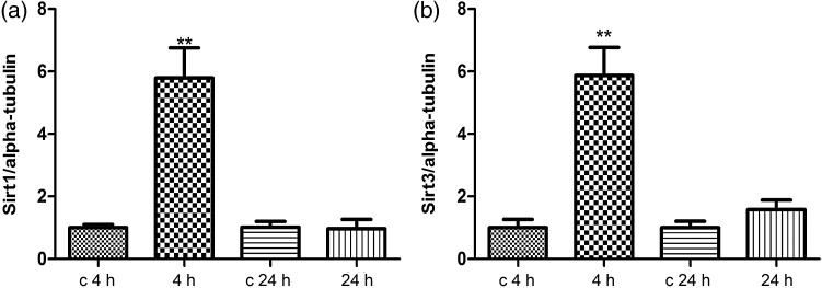
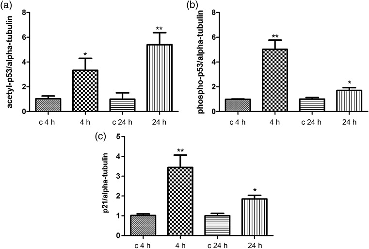
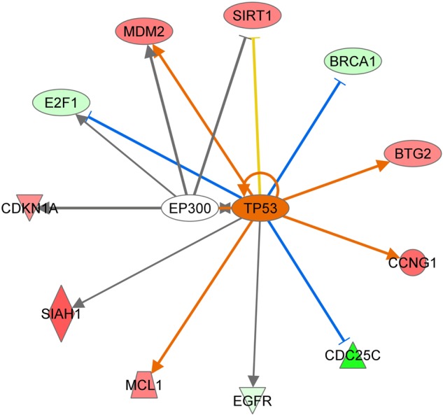
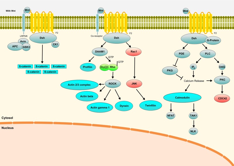
Reversible lysine acetylation is a highly regulated post-translational protein modification that is known to regulate several signaling pathways. However, little is known about the radiation-induced changes in the acetylome. In this study, we analyzed the acute post-translational acetylation changes in primary human cardiac microvascular endothelial cells 4 h after a gamma radiation dose of 2 Gy. The acetylated peptides were enriched using anti-acetyl conjugated agarose beads. A total of 54 proteins were found to be altered in their acetylation status, 23 of which were deacetylated and 31 acetylated. Pathway analyses showed three protein categories particularly affected by radiation-induced changes in the acetylation status: the proteins involved in the translation process, the proteins of stress response, and mitochondrial proteins. The activation of the canonical and non-canonical Wnt signaling pathways affecting actin cytoskeleton signaling and cell cycle progression was predicted. The protein expression levels of two nicotinamide adenine dinucleotide (NAD+)-dependent deacetylases, sirtuin 1 and sirtuin 3, were significantly but transiently upregulated 4 but not 24 h after irradiation. The status of the p53 protein, a target of sirtuin 1, was found to be rapidly stabilized by acetylation after radiation exposure. These findings indicate that post-translational modification of proteins by acetylation and deacetylation is essentially affecting the radiation response of the endothelium.

Keywords: acetylation; endothelial cell; heart; ionizing radiation; proteomics; sirtuins.


Figures

Fig. 1.
Fig. 1.
Pathway analysis of radiation-induced acetylated and deacetylated proteins. All differentially regulated acetylated and deacetylated proteins were imported into the GeneMANIA and IPA software tools. (a) GeneMANIA identified translational initiation as the most significant protein class affected by irradiation on the acetylation level. The striped brown and gray circles represent the identified acetylated/deacetylated proteins, whereas the uniquely colored circles represent the predicted interactive proteins. The brown color shows the network involved in protein synthesis; the gray color indicates proteins only indirectly related to protein synthesis. (b) The most affected biological networks from the IPA analysis were cell death and survival, cellular movement, cellular development, cellular morphology and protein trafficking; these are shown as a merged network. The gray molecules represent proteins found to be altered in their acetylation status in this study; the white molecules are interaction partners predicted by the software. The solid arrows and lines represent direct interactions, and the dotted arrows and lines indirect interactions.
Fig. 2.
Fig. 2.
Immunoblot analysis of the SIRT1 and SIRT3 expression. The extracted proteins from control and irradiated cells at 4 or 24 h post-irradiation were separated by 12% SDS-PAGE and analyzed by immunoblotting with antibodies against (a) SIRT1; (b) SIRT3. The columns represent the average ratios with standard errors of the mean (SEMs) of relative protein expression in sham-irradiated and irradiated cells. The protein bands (E) were quantified using ImageQuant 5.2 software (GE Healthcare) by integration of all the pixel values in the band area after background correction and normalized to alpha-tubulin expression. Three biological replicates were used for each experiment. *P ≤ 0.05; **P ≤ 0.01 (Student's t-test).
Fig. 3.
Fig. 3.
Immunoblot analysis of the radiation-induced changes in the p53 acetylation and phosphorylation status and in the p21 expression level. The extracted proteins from control and irradiated cells at 4 or 24 h after exposure were separated by 12% SDS-PAGE and analyzed by immunoblotting with antibodies against (a) anti-acetyl-p53; (b) anti-phospho-p53; (c) p21. The columns represent the average ratios with standard errors of the mean (SEMs) of relative protein expression/modification in sham- and irradiated cells. The protein bands (E) were quantified using ImageQuant 5.2 software (GE Healthcare) by integration of all the pixel values in the band area after background correction and normalized to the alpha-tubulin (αTub) expression. Three biological replicates were used for each experiment. *P ≤ 0.05; **P ≤ 0.01 (Student's t-test).
Fig. 4.
Fig. 4.
p53-related deregulated target genes using IPA software tool. The predicted interactions between TP53 (p53) and its target genes are shown by arrow (activation) or blocked arrow (inactivation). SIRT1 = sirtuin 1, CDC25C = cell division cycle 25C, EGFR = epidermal growth factor receptor, MCL1 = myeloid cell leukemia 1, SIAH1 = E3 ubiquitin-protein ligase 1; CDKNA1 = cyclin-dependent kinase inhibitor 1A, E2F1 = transcription factor E2F1, MDM2 = E3 ubiquitin-protein ligase, BRCA1 = breast cancer 1, BTG2 = BTG family member 2, anti-proliferative protein, CCNG1 = cyclin G1, EP300 = E1A-binding protein p300. Ingenuity upstream analysis predicted activation of the p53 gene by 2.156 activation score and overlap P-value of 5,39E10. Given the observed differential regulation of a gene (‘up’ or ‘down’) in the dataset, the activation state of an upstream regulator is determined by the regulation direction associated with the relationship from the regulator to the gene. The overlap P-value measures whether there is a statistically significant overlap between the dataset genes and the genes that are regulated by a transcriptional regulator. It is calculated using Fisher's Exact Test, and significance is generally attributed to P-values < 0.01.
Fig. 5.
Fig. 5.
Wnt canonical and non-canonical pathways affected 4 h after irradiation in HCMEC. Blue color represents proteins regulated by acetylation; green color represents proteins regulated at the level of total expression in endothelial cells [25, 73]; and red color represents proteins predicted activated using bioinformatics tools. Gray color represents proteins not found to be deregulated after irradiation.

References

    1. Choudhary C, Kumar C, Gnad F, et al. Lysine acetylation targets protein complexes and co-regulates major cellular functions. Science 2009;325:834–40. - PubMed
    1. Lee KK, Workman JL. Histone acetyltransferase complexes: one size doesn't fit all. Nat Rev Mol Cell Biol 2007;8:284–95. - PubMed
    1. Shahbazian MD, Grunstein M. Functions of site-specific histone acetylation and deacetylation. Annu Rev Biochem 2007;76:75–100. - PubMed
    1. Frye RA. Phylogenetic classification of prokaryotic and eukaryotic Sir2-like proteins. Biochem Biophys Res Commun 2000;273:793–8. - PubMed
    1. Morris BJ. Seven sirtuins for seven deadly diseases of aging. Free Radic Biol Med 2013;56:133–71. - PubMed

Publication types