Shape variation and covariation of upper and lower dental arches of an orthodontic population
- PMID: 25840587
- PMCID: PMC4914756
- DOI: 10.1093/ejo/cjv019
Shape variation and covariation of upper and lower dental arches of an orthodontic population
Abstract
Objectives: This study aimed to quantify the patterns of shape variability and the extent and patterns of shape covariation between the upper and lower dental arch in an orthodontic population.
Methods: Dental casts of 133 white subjects (61 males, 72 females; ages 10.6-26.6) were scanned and digitized in three dimensions. Landmarks were placed on the incisal margins and on the cusps of canines, premolars, and molars. Geometric morphometric methods were applied (Procrustes superimposition and principal component analysis). Sexual dimorphism and allometry were evaluated with permutation tests and age-size and age-shape correlations were computed. Two-block partial least squares analysis was used to assess covariation of shape.
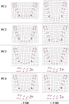
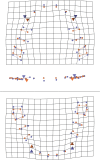
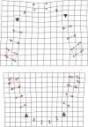
Results: The first four principal components represented shape patterns that are often encountered and recognized in clinical practice, accounting for 6-31 per cent of total variance. No shape sexual dimorphism was found, nevertheless, there was statistically significant size difference between males and females. Allometry was statistically significant, but low (upper: R(2) = 0.0528, P < 0.000, lower: R (2) = 0.0587, P < 0.000). Age and shape were weakly correlated (upper: R(2) = 0.0370, P = 0.0001, lower: R (2) = 0.0587, P = 0.0046). Upper and lower arches covaried significantly (RV coefficient: 33 per cent). The main pattern of covariation between the dental arches was arch width (80 per cent of total covariance); the second component related the maxillary canine vertical position to the mandibular canine labiolingual position (11 per cent of total covariance).
Limitations: Results may not be applicable to the general population. Age range was wide and age-related findings are limited by the cross-sectional design. Aetiology of malocclusion was also not considered.
Conclusions: Covariation patterns showed that the dental arches were integrated in width and depth. Integration in the vertical dimension was weak, mainly restricted to maxillary canine position.
© The Author 2015. Published by Oxford University Press on behalf of the European Orthodontic Society. All rights reserved. For permissions, please email: journals.permissions@oup.com.
Figures


References
-
- Brader A.C. (1972) Dental arch form related with intraoral forces: PR=C. American Journal of Orthodontics, 61, 541–561. - PubMed
-
- Harris E.F. (1997) A longitudinal study of arch size and form in untreated adults. American Journal of Orthodontics and Dentofacial Orthopedics, 111, 419–427. - PubMed
-
- Braun S., Hnat W.P., Fender D.E., Legan H.L. (1998) The form of the human dental arch. The Angle Orthodontist, 68, 29–36. - PubMed
-
- Raberin M., Laumon B., Martin J.L., Brunner F. (1993) Dimensions and form of dental arches in subjects with normal occlusions. American Journal of Orthodontics and Dentofacial Orthopedics, 104, 67–72. - PubMed
-
- Lee R.T. (1999) Arch width and form: a review. American Journal of Orthodontics and Dentofacial Orthopedics, 115, 305–313. - PubMed
MeSH terms
LinkOut - more resources
Full Text Sources
Other Literature Sources

