

Outer membrane vesicles - offensive weapons or good Samaritans?
- PMID: 25840612
- PMCID: PMC4385126
- DOI: 10.3402/jom.v7.27468
Outer membrane vesicles - offensive weapons or good Samaritans?
Abstract
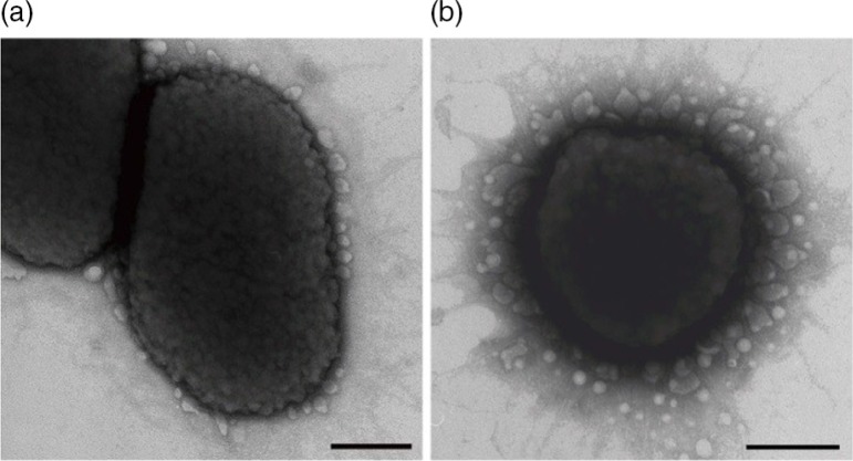
Outer membrane vesicles (OMVs) from Gram-negative bacteria were first considered as artifacts and were followed with disbelief and bad reputation. Later, their existence was accepted and they became characterized as bacterial bombs, virulence bullets, and even decoys. Today, we know that OMVs also can be involved in cell-cell signaling/communication and be mediators of immune regulation and cause disease protection. Furthermore, OMVs represent a distinct bacterial secretion pathway selecting and protecting their cargo, and they can even be good Samaritans providing nutrients to the gut microbiota maintaining commensal homeostasis beneficial to the host. The versatility in functions of these nanostructures is remarkable and includes both defense and offense. The broad spectrum of usability does not stop with that, as it now seems that OMVs can be used as vaccines and adjuvants or vehicles engineered for drug treatment of emerging and new diseases not only caused by bacteria but also by virus. They may even represent new ways of selective drug treatment.
Keywords: defense; offense; outer membrane vesicles; versatility in functions.
Figures
Similar articles
-
Bacterial Outer Membrane Vesicles: Role in Pathogenesis and Host-Cell Interactions.Antibiotics (Basel). 2023 Dec 28;13(1):32. doi: 10.3390/antibiotics13010032. Antibiotics (Basel). 2023. PMID: 38247591 Free PMC article. Review.
-
Activation of Immune and Defense Responses in the Intestinal Mucosa by Outer Membrane Vesicles of Commensal and Probiotic Escherichia coli Strains.Front Microbiol. 2016 May 11;7:705. doi: 10.3389/fmicb.2016.00705. eCollection 2016. Front Microbiol. 2016. PMID: 27242727 Free PMC article.
-
Outer membrane vesicles function as offensive weapons in host-parasite interactions.Microbes Infect. 2010 Oct;12(11):791-8. doi: 10.1016/j.micinf.2010.05.008. Epub 2010 Jun 2. Microbes Infect. 2010. PMID: 20685339 Review.
-
Outer membrane vesicles engineered to express membrane-bound antigen program dendritic cells for cross-presentation to CD8+ T cells.Acta Biomater. 2019 Jun;91:248-257. doi: 10.1016/j.actbio.2019.04.033. Epub 2019 Apr 17. Acta Biomater. 2019. PMID: 31003032
-
Outer Membrane Vesicles (OMVs) of Gram-negative Bacteria: A Perspective Update.Front Microbiol. 2017 Jun 9;8:1053. doi: 10.3389/fmicb.2017.01053. eCollection 2017. Front Microbiol. 2017. PMID: 28649237 Free PMC article. Review.
Cited by
-
Host-derived extracellular vesicles for antimicrobial defense.Microlife. 2021 Apr 16;2:uqab003. doi: 10.1093/femsml/uqab003. eCollection 2021. Microlife. 2021. PMID: 37223251 Free PMC article.
-
Invasion of Porphyromonas gingivalis strains into vascular cells and tissue.J Oral Microbiol. 2015 Aug 31;7:28788. doi: 10.3402/jom.v7.28788. eCollection 2015. J Oral Microbiol. 2015. PMID: 26329158 Free PMC article.
-
Outer membrane vesicles blebbing contributes to B. vulgatus mpk-mediated immune response silencing.Gut Microbes. 2018 Jan 2;9(1):1-12. doi: 10.1080/19490976.2017.1344810. Epub 2017 Jul 13. Gut Microbes. 2018. PMID: 28686482 Free PMC article.
-
Bioengineering commensal bacteria-derived outer membrane vesicles for delivery of biologics to the gastrointestinal and respiratory tract.J Extracell Vesicles. 2019 Jun 24;8(1):1632100. doi: 10.1080/20013078.2019.1632100. eCollection 2019. J Extracell Vesicles. 2019. PMID: 31275534 Free PMC article.
-
Proteomics of Aggregatibacter actinomycetemcomitans Outer Membrane Vesicles.PLoS One. 2015 Sep 18;10(9):e0138591. doi: 10.1371/journal.pone.0138591. eCollection 2015. PLoS One. 2015. PMID: 26381655 Free PMC article.
References
-
- Kaback HR. Transport studies in bacterial membrane vesicles. Science. 1974;186:882–92. - PubMed
LinkOut - more resources
Full Text Sources
Other Literature Sources
Research Materials