

Oncostatin M promotes mucosal epithelial barrier dysfunction, and its expression is increased in patients with eosinophilic mucosal disease
- PMID: 25840724
- PMCID: PMC4562861
- DOI: 10.1016/j.jaci.2015.01.043
Oncostatin M promotes mucosal epithelial barrier dysfunction, and its expression is increased in patients with eosinophilic mucosal disease
Abstract
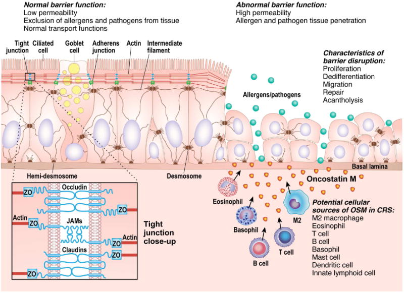
Background: Epithelial barrier dysfunction is thought to play a role in many mucosal diseases, including asthma, chronic rhinosinusitis (CRS), and eosinophilic esophagitis.
Objective: The objective of this study was to investigate the role of oncostatin M (OSM) in epithelial barrier dysfunction in human mucosal disease.
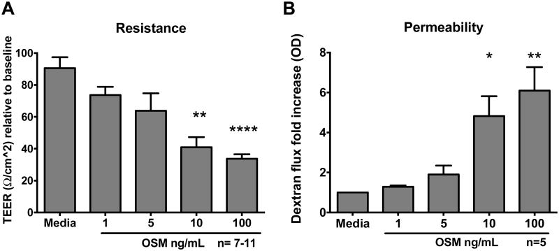
Methods: OSM expression was measured in tissue extracts, nasal secretions, and bronchoalveolar lavage fluid. The effects of OSM stimulation on barrier function of normal human bronchial epithelial cells and nasal epithelial cells cultured at the air-liquid interface were assessed by using transepithelial electrical resistance and fluorescein isothiocyanate-dextran flux. Dual-color immunofluorescence was used to evaluate the integrity of tight junction structures in cultured epithelial cells.
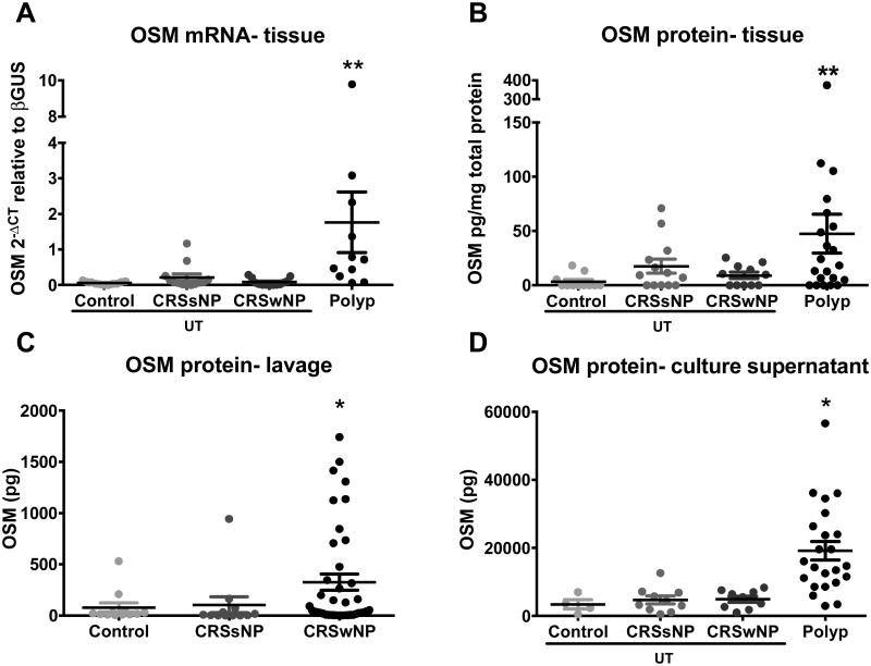
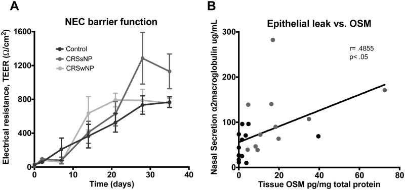
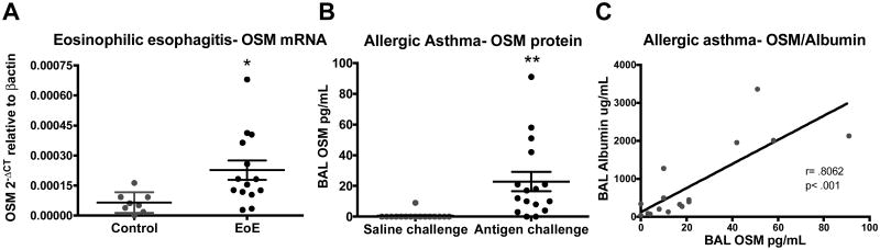
Results: Analysis of samples from patients with CRS showed that OSM mRNA and protein levels were highly increased in nasal polyps compared with those seen in control uncinate tissue (P < .05). OSM levels were also increased in bronchoalveolar lavage fluid of allergic asthmatic patients after segmental allergen challenge and in esophageal biopsy specimens from patients with eosinophilic esophagitis. OSM stimulation of air-liquid interface cultures resulted in reduced barrier function, as measured by decreased transepithelial electrical resistance and increased fluorescein isothiocyanate-dextran flux (P < .05). Alterations in barrier function by OSM were reversible, and the viability of epithelial cells was unaffected. OSM levels in lysates of nasal polyps and uncinate tissue positively correlated with levels of α2-macroglobulin, a marker of epithelial leak, in localized nasal secretions (r = 0.4855, P < .05).
Conclusions: These results suggest that OSM might play a role in epithelial barrier dysfunction in patients with CRS and other mucosal diseases.
Keywords: Oncostatin M; atopic asthma; chronic rhinosinusitis; eosinophilic esophagitis; epithelial barrier; tight junctions; transepithelial electrical resistance.
Copyright © 2015 American Academy of Allergy, Asthma & Immunology. Published by Elsevier Inc. All rights reserved.
Figures

References
-
- Podolsky DK. Mucosal immunity and inflammation. V. Innate mechanisms of mucosal defense and repair: the best offense is a good defense. The American journal of physiology. 1999;277(3 Pt 1):G495–499. - PubMed
-
- Fanning AS, Jameson BJ, Jesaitis LA, Anderson JM. The tight junction protein ZO-1 establishes a link between the transmembrane protein occludin and the actin cytoskeleton. The Journal of biological chemistry. 1998;273(45):29745–29753. - PubMed
-
- Oppenheim JJ, Tewary P, de la Rosa G, Yang D. Alarmins initiate host defense. Advances in experimental medicine and biology. 2007;601:185–194. - PubMed
-
- Trautmann A, Kruger K, Akdis M, Muller-Wening D, Akkaya A, Brocker EB, et al. Apoptosis and loss of adhesion of bronchial epithelial cells in asthma. International archives of allergy and immunology. 2005;138(2):142–150. - PubMed
-
- Holgate ST, Lackie P, Wilson S, Roche W, Davies D. Bronchial epithelium as a key regulator of airway allergen sensitization and remodeling in asthma. American journal of respiratory and critical care medicine. 2000;162(3 Pt 2):S113–117. - PubMed
Publication types
MeSH terms
Substances
Grants and funding
LinkOut - more resources
Full Text Sources
Other Literature Sources
Medical
