Review
. 2015 May;204(2):134-43.
doi: 10.1016/j.tvjl.2015.02.017. Epub 2015 Feb 26.

Porcine epidemic diarrhea virus infection: Etiology, epidemiology, pathogenesis and immunoprophylaxis

Affiliations
Review

Porcine epidemic diarrhea virus infection: Etiology, epidemiology, pathogenesis and immunoprophylaxis

Kwonil Jung et al. Vet J. 2015 May.

Abstract

Porcine epidemic diarrhea virus (PEDV), a member of the genera Alphacoronavirus in the family Coronaviridae, causes acute diarrhea/vomiting, dehydration and high mortality in seronegative neonatal piglets. For the last three decades, PEDV infection has resulted in significant economic losses in the European and Asian pig industries, but in 2013-2014 the disease was also reported in the US, Canada and Mexico. The PED epidemic in the US, from April 2013 to the present, has led to the loss of more than 10% of the US pig population. The disappearance and re-emergence of epidemic PED indicates that the virus is able to escape from current vaccination protocols, biosecurity and control systems. Endemic PED is a significant problem, which is exacerbated by the emergence (or potential importation) of multiple PEDV variants. Epidemic PEDV strains spread rapidly and cause a high number of pig deaths. These strains are highly enteropathogenic and acutely infect villous epithelial cells of the entire small and large intestines although the jejunum and ileum are the primary sites. PEDV infections cause acute, severe atrophic enteritis accompanied by viremia that leads to profound diarrhea and vomiting, followed by extensive dehydration, which is the major cause of death in nursing piglets. A comprehensive understanding of the pathogenic characteristics of epidemic or endemic PEDV strains is needed to prevent and control the disease in affected regions and to develop an effective vaccine. This review focuses on the etiology, epidemiology, disease mechanisms and pathogenesis as well as immunoprophylaxis against PEDV infection.

Keywords: Disease; Pathogenesis; Porcine epidemic diarrhea virus; Review; Swine; Virus.


Figures

Fig. 1
Fig. 1
Histopathology, localization of porcine epidemic diarrhea virus (PEDV) antigens by immunofluorescence staining, and apoptotic cells by an in situ TUNEL assay in the intestine of gnotobiotic pigs inoculated with US PEDV strain PC21A. (A) Hematoxylin and eosin (H&E)-stained jejunum of an inoculated pig at post-inoculation hour (PIH) 46 (at onset of clinical signs), showing acute diffuse, severe atrophic jejunitis with ratios of villous height to crypt depth (VH:CD) of ≤1. Original magnification ×200. (B) Immunofluorescent (IF) staining of jejunum of an inoculated pig at PIH 30 (4–5 h after onset of clinical signs), showing that the epithelial cells lining atrophied villi are positive for PEDV antigen. Original magnification ×200. (C) In situ TUNEL staining of formalin-fixed, paraffin-embedded jejunum of the inoculated pig (Panel B), showing no increase of TUNEL-positive (apoptotic) cells (red staining) in the epithelium lining atrophied villi positive for PEDV antigen by IF staining, compared to Panel D (negative control). Original magnification ×200. (D) In situ TUNEL staining of formalin-fixed, paraffin-embedded jejunum of a non-inoculated, negative control pig, showing few TUNEL-positive (apoptotic) cells (red staining) in the intestinal villous epithelium. Note a few TUNEL-positive cells (red staining) in the lamina propria. Original magnification ×200. IL, intestinal lumen. Nuclei were stained with blue-fluorescent 4′, 6-diamidino-2-phenylindole dihydrochloride in Panel B. TUNEL, in situ terminal deoxynucleotidyl transferase-mediated dUTP nick end labeling.
Fig. 2
Fig. 2
Decreased numbers of goblet cells in the small intestine of gnotobiotic pigs inoculated with US porcine epidemic diarrhea virus (PEDV) strain PC21A, as determined by toluidine blue staining. (A) Jejunum of an inoculated pig at post-inoculation hour (PIH) 72 (26–28 h after onset of clinical signs), showing few goblet cells per villus. Original magnification, ×200. (B) Jejunum of a negative control pig, showing small to moderate numbers of goblet cells (arrows) per villus. Original magnification, ×200. (C) Ileum of an inoculated pig at PIH 72 (26–28 h after onset of clinical signs), showing few goblet cells per villus. Original magnification, ×80. (D) Ileum of a negative control pig, showing moderate to large numbers of goblet cells (arrows) per villus. Original magnification, ×80.
Fig. 3
Fig. 3
Expression of the tight junction protein, zonula occludin (ZO)-1, (A, B) and adherens junction protein, E-cadherin, (C, D) by immunofluorescence staining in the small intestine of gnotobiotic pigs inoculated with US porcine epidemic diarrhea virus (PEDV) PC21A. (A) Jejunum of a negative control pig, showing well-organized distribution and extensive expression of ZO-1 (green staining) on the apical surface of villous epithelial cells. Original magnification, ×400. (B) Jejunum of an inoculated pig at post-inoculation hour (PIH) 30 (4–5 h after onset of clinical signs), showing disorganized, irregular distribution and moderately reduced expression of ZO-1 (green staining), compared to the negative control counterpart (Panel A), on the apical surface of villous epithelial cells. Original magnification, ×400. (C) Jejunum of a negative control pig, showing well-organized distribution and extensive expression of E-cadherin (green staining) on the apical and basolateral surface of villous epithelial cells and also mildly in the cytoplasm. Original magnification, ×600. (D) Jejunum of an inoculated pig at PIH 30 (4–5 h after onset of clinical signs), showing disorganized, irregular distribution and moderately decreased expression of E-cadherin (green staining) on the apical and basolateral surface of villous epithelial cells, compared to the negative control counterpart (Panel C). Original magnification, ×600. Nuclei were stained with blue-fluorescent 4′, 6-diamidino-2-phenylindole dihydrochloride. Immunofluorescence staining using monoclonal antibodies against human recombinant ZO-1 and E-cadherin (Invitrogen).
Fig. 4
Fig. 4
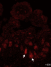
Localization of LGR5+ crypt stem cells by immunofluorescence staining in the small intestine of gnotobiotic pigs inoculated with US porcine epidemic diarrhea virus (PEDV) strain PC21A. Jejunum of a PEDV-inoculated pig at post-inoculation hour 30 (4–5 h after onset of clinical signs), showing large numbers of LGR5+ crypt stem cells (red staining; arrows) in the crypt cell layer of atrophied villi. Original magnification, ×300. Immunofluorescence staining using a polyclonal antibody against human LGR5 (Novus Biologicals). LGR5+ crypt stem cells, LGR5 (leucine-rich repeat-containing G protein-coupled receptor 5)-positive crypt base columnar cells.
Fig. 5
Fig. 5
Localization of CD4 or CD8-positive T cells by immunofluorescence staining in the small intestine of gnotobiotic pigs inoculated with US porcine epidemic diarrhea virus (PEDV) strain PC21A. (A, B) Jejunum of an inoculated pig at 120 h after onset of clinical signs, showing low to moderate numbers of CD4+ (A) and CD8+ (B) T cells (arrows) in the lamina propria. Original magnification, all ×400. (C) Jejunum of a non-inoculated, negative control pig, showing no detectable CD4+ T cells in the intestinal section. Original magnification, ×200. Nuclei were stained with blue-fluorescent 4′, 6-diamidino-2-phenylindole dihydrochloride.

Comment in

Similar articles

Cited by

References

    1. Alonso C., Goede D.P., Morrison R.B., Davies P.R., Rovira A., Marthaler D.G., Torremorell M. Evidence of infectivity of airborne porcine epidemic diarrhea virus and detection of airborne viral RNA at long distances from infected herds. Veterinary Research. 2014;45:73. - PMC - PubMed
    1. Bohl E.H., Kohler E.M., Saif L.J., Cross R.F., Agnes A.G., Theil K.W. Rotavirus as a cause of diarrhea in pigs. Journal of the American Veterinary Medical Association. 1978;172:458–463. - PubMed
    1. Bosch B.J., van der Zee R., de Haan C.A., Rottier P.J. The coronavirus spike protein is a class I virus fusion protein: Structural and functional characterization of the fusion core complex. Journal of Virology. 2003;77:8801–8811. - PMC - PubMed
    1. Burkey T.E., Skjolaas K.A., Minton J.E. Porcine mucosal immunity of the gastrointestinal tract. Journal of Animal Science. 2009;87:1493–1501. - PubMed
    1. Carvajal A., Lanza I., Diego R., Rubio P., Carmenes P. Evaluation of a blocking ELISA using monoclonal antibodies for the detection of porcine epidemic diarrhea virus and its antibodies. Journal of Veterinary Diagnostic Investigation. 1995;7:60–64. - PubMed

Publication types

MeSH terms